FIGURE SUMMARY
Title

Estrogen accelerates heart regeneration by promoting the inflammatory response in zebrafish

Authors
Xu, S., Xie, F., Tian, L., Fallah, S., Babaei, F., Manno, S.H., Manno Iii, F.A.M., Zhu, L., Wong, K.F., Liang, Y., Ramalingam, R., Sun, L., Wang, X., Plumb, R., Gethings, L., Lam, Y.W., Cheng, S.H.
Source
Full text @ J. Endocrinol.

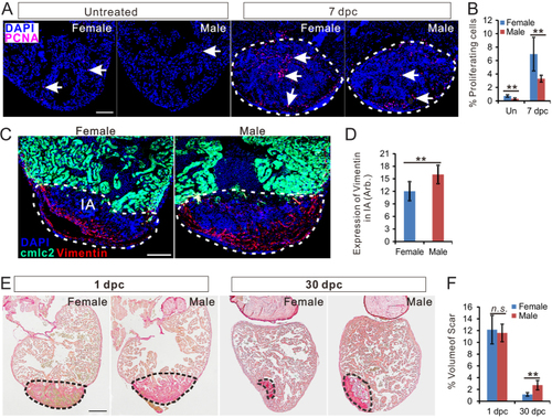
Zebrafish heart regeneration is sexually dimorphic. (A) PCNA immunofluorescence (red) in the heart of untreated female, untreated male, 7 dpc female (c) and 7 dpc male. Scale bars: 100μm. (B) Quantification of percentage of PCNA-positive cells (mean ± s.d., n = 7) in panel A. (C) Vimentin immunofluorescence (red) in female and male Tg (cmlc2: eGFP, green) zebrafish hearts at 7 dpc. Scale bars: 100 μm. (D) Quantification of vimentin expression in the injured area of female and male zebrafish heart (marked by dashed lines, mean ± s.d., n = 8). (E) Picrosirus red staining of female and male zebrafish hearts at 1 dpc and 30 dpc. (F) Quantification of scar volume (marked by dashed lines, mean ± s.d., n = 9~12) between females and male zebrafish. Scale bars: 200 μm. Two-tail t test in figure B, D and F, **P < 0.01, n.s., not significant. Un, untreated; Dpc, days post-cryoinjury; CI, Cryoinjury.

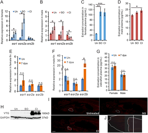
Cardiac damage increased estrogen expression in zebrafish. (A, B, C, D, E, F and G) qRT-PCR showing the expression of estrogen receptor genes in the heart (A, B) and fin (E, F) of zebrafish 7 days after CI; and plasma E2 concentration in zebrafish with heart (C, D) or fin (G) injury at 7 days. n = 3, two-tail t test (A, B, E, F, G), and one-way ANOVA with LSD post hoc test (C, D), *P < 0.05, **P < 0.01, n.s, not significant. (H) Detection of vitellogenin by Western blotting of plasma collected from three independent untreated male zebrafish and fish one day after SO and CI. (I) Detection of VTG (red) in untreated male zebrafish and in male zebrafish on day 7 after heart CI and SO. Scale bars: 1 mm. (J) Detection of VTG in uninjured caudal fin and on 7 days after fin amputation. The dashed white line showed the shape of fin, and the white line showed the amputation site. Scale bar: 1 mm. Un, untreated; SO, sham-operation; and CI, cryoinjury.

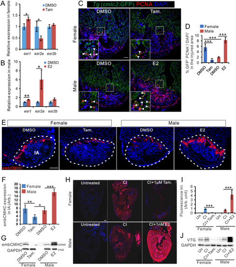
Estrogen promotes regeneration in zebrafish heart. (A and B) qRT-PCR showing the expression of estrogen receptor genes in the heart of female fish (A) and male zebrafish (B) with DMSO, tamoxifen or E2 treatment at 7 days after cryoinjury. n = 3. (C) PCNA immunofluorescence (red) in the heart of male Tg (cmlc2: eGFP) zebrafish exposed to 1 nM E2 for 7 days after CI. Scale bar: 100 μm. (D). Quantification of proliferating cardiomyocytes in panel C (mean ± s.d., n = 5). One-way ANOVA with LSD post hoc test, *P = 0.05, ***P < 0.001. (E) embCMHC immunofluorescence (red) in the heart of untreated females, females treated with 1 μM tamoxifen (Tam.), untreated males, and males treated with 1 nM E2 for 7 days after CI. Scale bar: 200μm. (F) Quantification of embCMHC staining in panel E (mean ± s.d., n = 4~5) in the injured area. One-way ANOVA with LSD post hoc test, *P = 0.05, **P = 0.01, ***P < 0.001. (G) Expression of embCMHC in the sample shown in E, as detected by Western blotting. (H) Vitellogenin (VTG) immunofluorescence (red) in the heart of untreated females, 7 dpc females, 7 dpc females treated with 1 μM Tamoxifen, untreated males, 7 dpc males, and 7 dpc males treated with 1nM E2. Scale bar: 100 μm. (I) Quantification of VTG staining (mean ± s.d., n = 4~5) in panel H. One-way ANOVA with LSD post hoc test, ***P < 0.001. (J) Expression of VTG in samples shown in panel H, as detected by Western blotting. Un, untreated, SO, sham-operation, and CI, cryoinjury.

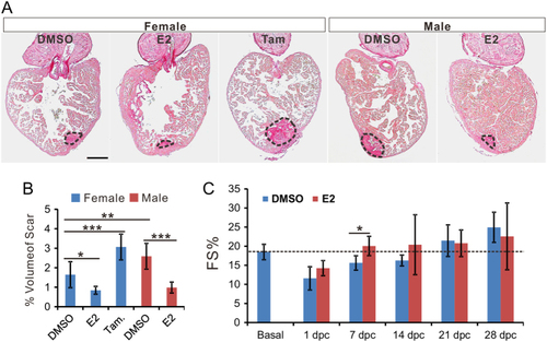
Estrogen accelerates scar reduction and promotes recovery of cardiac function. (A) Picrosirus red staining of the heart from female zebrafish treated with DMSO, E2 (1 nM) and tamoxifen (1 μM); and male zebrafish treated with DMSO and E2 (1 nM) at 30 dpc. Scale bar: 200 μm. (B) Quantification of scar volume from samples in panel A (marked by dash lines, mean ± s.d., n = 5~6). One-way ANOVA with LSD post hoc test, *P < 0.05, **P < 0.01, and ***P < 0.001. (C) Fractional shortening (FS) measured by echocardiography of male zebrafish before (basal) and at different time points during recovery in DMSO or E2 (1 nM) after CI. Two-way ANOVA with LSD post hoc test, *P < 0.05, n = 5~6.

Estrogen induces inflammation in the injured zebrafish heart. A. Gene Ontogeny (GO) terms significantly enriched in genes differentially expressed in female vs male hearts at 7 dpc, n = 3. (B) Comparison of gene expression patterns at 7 dpc in the hearts of female vs male, E2 (1 nM)-treated male vs untreated male, and tamoxifen (1 μM)-treated female vs untreated female, n = 3. (C) Expression of immune and inflammation-related genes in the datasets, n = 3. (D, E and F) Expression of ifn-γ at 7dpc in female and male zebrafish heart and uninjured fish (D). Tamoxifen decreased (E) and E2 increased (F) the expression of ifn-γ in females and males at 7 dpc. n = 4, two-tail t test, *P < 0.05, **P < 0.01, ***P < 0.001. (G and H) Western blot of STAT1 and STAT3 in female and male heart at 7 dpc using different treatments. H, bar chart showing the quantification of STAT1 expression in male fish in panel G, n = 3, two-tail t test, *P < 0.05.

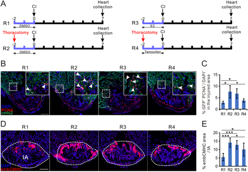
Estrogen preconditioning treatment promotes zebrafish heart regenerative process. (A) Experimental design of data presented in this figure. (B) PCNA immunofluorescence (red) in the heart of male Tg (cmlc2: eGFP) zebrafish heart from regimens R1-R4. Arrowheads in the white line bounded area indicate proliferating cardiomyocytes (PCNA+/GFP+). Scale bar: 100 μm. (C) Quantification of proliferating cardiomyocytes (PCNA+/GFP+) in panel B (mean ± s.d., n = 4~5). One-way ANOVA with LSD post hoc test, *P < 0.05. (D) embCMHC immunofluorescence (red) in the male heart from regimens R1-R4. (E) Quantification of embCMHC staining in panel E (mean ± s.d., n = 5~6) in the injured area. Scale bars: 100 μm. One-way ANOVA with LSD post hoc test, *P = 0.05, ***P < 0.001.

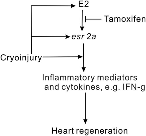
Proposed model estrogen role in zebrafish heart regeneration. Heart cryoinjury triggers the inflammation response and plasma estrogen upregulation. The increase of plasma estrogen induces expression of esr2a, which can be inhibited by tamoxifen. Estrogen promotes heart regeneration through esr2a induced and injury directly induced inflammation response.

Acknowledgments
This image is the copyrighted work of the attributed author or publisher, and ZFIN has permission only to display this image to its users. Additional permissions should be obtained from the applicable author or publisher of the image. Full text @ J. Endocrinol.