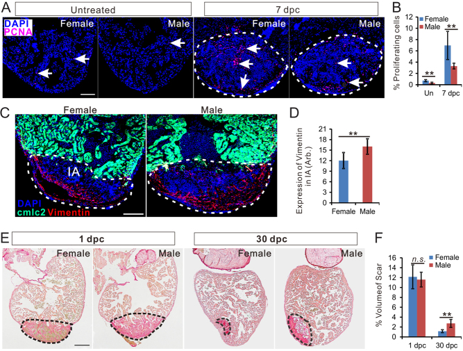
IMAGE

Figure 1

ID
ZDB-IMAGE-200229-11
Source
Figures for Xu et al., 2020
Image
Figure Caption

Figure 1

Zebrafish heart regeneration is sexually dimorphic. (A) PCNA immunofluorescence (red) in the heart of untreated female, untreated male, 7 dpc female (c) and 7 dpc male. Scale bars: 100μm. (B) Quantification of percentage of PCNA-positive cells (mean ± s.d., n = 7) in panel A. (C) Vimentin immunofluorescence (red) in female and male Tg (cmlc2: eGFP, green) zebrafish hearts at 7 dpc. Scale bars: 100 μm. (D) Quantification of vimentin expression in the injured area of female and male zebrafish heart (marked by dashed lines, mean ± s.d., n = 8). (E) Picrosirus red staining of female and male zebrafish hearts at 1 dpc and 30 dpc. (F) Quantification of scar volume (marked by dashed lines, mean ± s.d., n = 9~12) between females and male zebrafish. Scale bars: 200 μm. Two-tail t test in figure B, D and F, **P < 0.01, n.s., not significant. Un, untreated; Dpc, days post-cryoinjury; CI, Cryoinjury.

Acknowledgments
This image is the copyrighted work of the attributed author or publisher, and ZFIN has permission only to display this image to its users. Additional permissions should be obtained from the applicable author or publisher of the image. Full text @ J. Endocrinol.