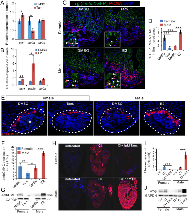
IMAGE

Figure 3

ID
ZDB-IMAGE-200229-13
Source
Figures for Xu et al., 2020
Image
Figure Caption

Figure 3

Estrogen promotes regeneration in zebrafish heart. (A and B) qRT-PCR showing the expression of estrogen receptor genes in the heart of female fish (A) and male zebrafish (B) with DMSO, tamoxifen or E2 treatment at 7 days after cryoinjury. n = 3. (C) PCNA immunofluorescence (red) in the heart of male Tg (cmlc2: eGFP) zebrafish exposed to 1 nM E2 for 7 days after CI. Scale bar: 100 μm. (D). Quantification of proliferating cardiomyocytes in panel C (mean ± s.d., n = 5). One-way ANOVA with LSD post hoc test, *P = 0.05, ***P < 0.001. (E) embCMHC immunofluorescence (red) in the heart of untreated females, females treated with 1 μM tamoxifen (Tam.), untreated males, and males treated with 1 nM E2 for 7 days after CI. Scale bar: 200μm. (F) Quantification of embCMHC staining in panel E (mean ± s.d., n = 4~5) in the injured area. One-way ANOVA with LSD post hoc test, *P = 0.05, **P = 0.01, ***P < 0.001. (G) Expression of embCMHC in the sample shown in E, as detected by Western blotting. (H) Vitellogenin (VTG) immunofluorescence (red) in the heart of untreated females, 7 dpc females, 7 dpc females treated with 1 μM Tamoxifen, untreated males, 7 dpc males, and 7 dpc males treated with 1nM E2. Scale bar: 100 μm. (I) Quantification of VTG staining (mean ± s.d., n = 4~5) in panel H. One-way ANOVA with LSD post hoc test, ***P < 0.001. (J) Expression of VTG in samples shown in panel H, as detected by Western blotting. Un, untreated, SO, sham-operation, and CI, cryoinjury.

Acknowledgments
This image is the copyrighted work of the attributed author or publisher, and ZFIN has permission only to display this image to its users. Additional permissions should be obtained from the applicable author or publisher of the image. Full text @ J. Endocrinol.