
- Title
-
Cell senescence contributes to tissue regeneration in zebrafish
- Authors
- Da Silva-Álvarez, S., Guerra-Varela, J., Sobrido-Cameán, D., Quelle, A., Barreiro-Iglesias, A., Sánchez, L., Collado, M.
- Source
- Full text @ Aging Cell
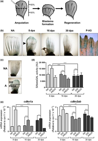
Pectoral fin amputation induces features of cell senescence. (a) Schematic representation of the fin amputation system used throughout the study. (b) Representative photomicrographs of fins stained for SAbetaGal or phospho‐histone 3 (P‐H3, right panel) after amputations (NA: nonamputated; 8, 16, and 30 dpa: days postamputation). Co‐staining of P‐H3 was done at 8 dpa. Arrowhead shows the amputation plane. (c) Schematic representation showing the different types of samples used in the study (NA: nonamputated; A: amputated; DIS: distal area; PROX: proximal). (d) SAbetaGal activity measured using Galacton substrate after 8, 16, and 30 days postamputation (dpa) (from 5–10 animals per condition). (e) Expression levels by QPCR of |
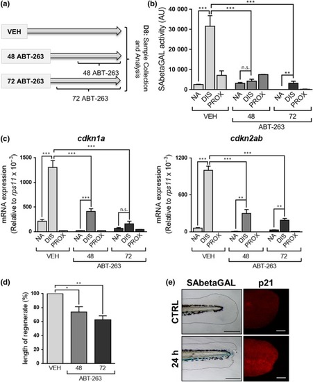
Removal of senescent cells impairs fin regeneration. (a) Schematic representation of the experimental strategy followed to analyze the effect of removing senescent cells from amputated fins after incubation with ABT‐263 for 48 or 72 hr, or treated with vehicle (VEH). (b) SAbetaGal activity measured using Galacton substrate at 8 days postamputation and after treatment with ABT‐263 for 48 or 72 hr, or with vehicle (VEH) (from 5–10 animals per condition). (c) Expression levels by QPCR of |