
|
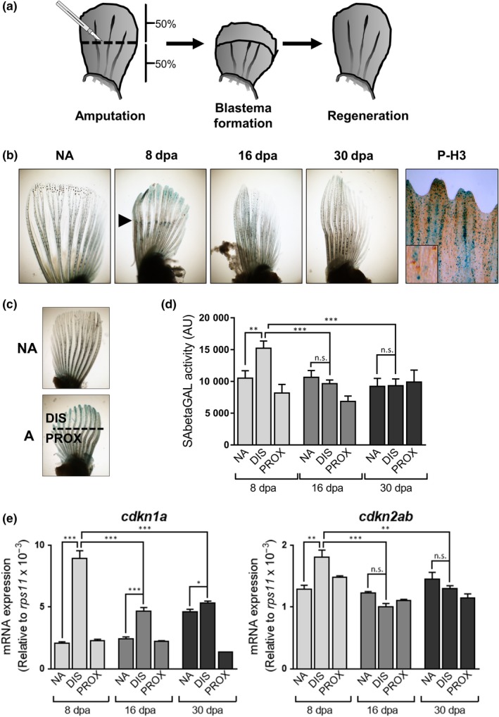
Figure 1
Pectoral fin amputation induces features of cell senescence. (a) Schematic representation of the fin amputation system used throughout the study. (b) Representative photomicrographs of fins stained for SAbetaGal or phospho‐histone 3 (P‐H3, right panel) after amputations (NA: nonamputated; 8, 16, and 30 dpa: days postamputation). Co‐staining of P‐H3 was done at 8 dpa. Arrowhead shows the amputation plane. (c) Schematic representation showing the different types of samples used in the study (NA: nonamputated; A: amputated; DIS: distal area; PROX: proximal). (d) SAbetaGal activity measured using Galacton substrate after 8, 16, and 30 days postamputation (dpa) (from 5–10 animals per condition). (e) Expression levels by QPCR of