FIGURE SUMMARY
Title

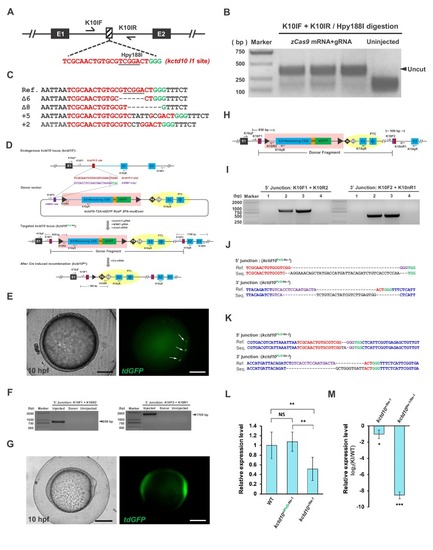
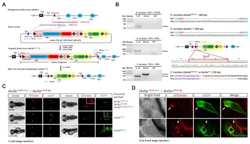
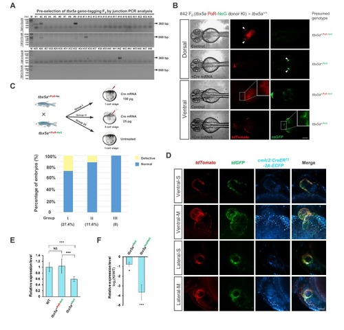
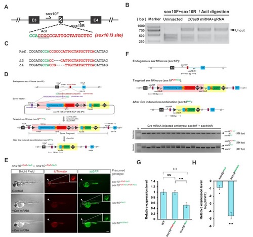
One-step efficient generation of dual-function conditional knockout and geno-tagging alleles in zebrafish

Authors
Li, W., Zhang, Y., Han, B., Li, L., Li, M., Lu, X., Chen, C., Lu, M., Zhang, Y., Jia, X., Zhu, Z., Tong, X., Zhang, B.
Source
Full text @ Elife

Evaluation of the expression of <italic>tbx5a</italic> and targeted insertion of the CKO + gene labeling PoNe donor at the <italic>tbx5a I2</italic> target site in founder embryos.

(A) The position and sequence of the tbx5a intron 2 (I2) target site designed for the Cas9/gRNA system. The protospacer sequence is shown in red, and the PAM is shown in green. (B) Indel efficiency evaluated by PCR and BtgI restriction endonuclease digestion. (C) Sequencing results of the uncut PCR products (corresponding to indel mutations) from B after cloning. (D) Mosaic expression of tdTomato in the heart (white arrowhead) and fin (white arrows) in a founder embryo after the injection of gRNAs purified by LiCl precipitation together with zCas9 mRNA and the tbx5a PoR-Ne donor from Figure 1A. Scale bar, 200 μm. (E) Junction PCR to detect NHEJ-mediated knockin events in founder embryos. The expected products (870 bp and 570 bp) were obtained by amplification with the corresponding primers shown in Figure 1A. Injected: Donor+Cas9/gRNA-injected embryos. Donor: tbx5a PoR-Ne donor plasmid. Uninjected: Uninjected embryos. (F) The expression of tbx5a in the zebrafish heart shown by whole-mount in situ hybridization (ventral view). The dotted lines denote the outline of the heart. Scale bar, 100 μm.

Evaluation of the indel efficiency of the <italic>tbx5a E3</italic> target site and phenotype analysis of the <italic>tbx5a</italic> indel mutation.

(A) The position and sequence of the tbx5a exon 3 (E3) target site designed for the Cas9/gRNA system. The protospacer sequence is shown in red, and the PAM is shown in green. (B) Targeting efficiency evaluated by PCR and AluI restriction endonuclease digestion. The result indicates that the indel efficiency is nearly 90%. (C) Sequencing results of the uncut PCR products (corresponding to indel mutations) from B after cloning. (D) Approximately 25% of embryos from the incross of tbx5a+/Δ5 heterozygotes showed defects in heart (black arrows) and pectoral fins (black arrowheads). Genotyping results revealed that all the defective embryos were tbx5aΔ5/Δ5 homozygotes (lower panel), while the siblings showed a normal morphology. The Tg(cmlc2:EGFP) transgenic background was introduced to reveal the heart morphology, and all the defective embryos also showed failure of cardiac looping. The dotted lines denote the outline of the heart. Scale bar, 200 μm. (E) qRT-PCR results showing the transcription level of the tbx5a locus in wild-type (WT) and tbx5a PoR-Ne donor KI zebrafish embryos at 72 hpf, using T5qF and T5qR primers. The tbx5a+/Ne and tbx5a+/PoR-Ne embryos were obtained from the crossing of the tbx5a PoR-Ne/PoR-Ne homozygotes with wild-type zebrafish with or without injection of Cre mRNA, respectively. The average expression level of wild-type embryos was set as 1. (F) qRT-PCR results using T5qF and T5qR primers, showing the transcription level of the tbx5a locus in the tbx5a+/Ne and tbx5aNe/Ne embryos derived from the Cre mRNA-injected tbx5a+/PoR-Ne and tbx5aPoR-Ne/PoR-Ne embryos, respectively. The original embryos were obtained from the crossing of tbx5aPoR-Ne/PoR-Ne homozygotes with tbx5a+/PoR-Ne heterozygote zebrafish. The expression levels in the KI embryos were normalized to the WT ones. Data are presented as the mean ±s.d., and a two-tailed Student’s t-test was applied to calculate p values in all the experiments. *: p<0.05. ***: p<0.001. NS: Not significant.

Strategy and evaluation of the targeted insertion of the PoG-Ne donor at the <italic>kctd10</italic> locus.

(A) The position and sequence of the kctd10 intron 1 (I1) target site designed for the Cas9/gRNA system. The protospacer sequence is shown in red, and the PAM is shown in green. (B) Targeting efficiency evaluated by PCR and Hpy188I restriction endonuclease digestion. (C) Sequencing results of the uncut PCR products (corresponding to indel mutations) from B after cloning. (D) Schematic diagram of the kctd10-2A-td GFP floxP 2PA-mutExon PoNe donor (abbreviated as kctd10 PoG-Ne donor) and the strategy of targeted insertion and conditional knockout using the CRISPR/Cas system. Primers K10qF and K10qR are used for qRT-PCR in L and M. (E) Images of a 10 hpf F0 zebrafish embryo after the injection of the kctd10 PoG-Ne donor together with zCas9 mRNA and corresponding gRNAs. White arrows indicate tdGFP signals. Scale bar, 200 μm. (F) Junction PCR to detect NHEJ-mediated knockin events in the injected founder embryos. Injected: Donor+Cas9/gRNA-injected embryos. Donor: kctd10 PoG-Ne donor plasmid. Uninjected: Uninjected embryos. (G) Images of a 10 hpf F1 zebrafish embryo from an outcross of the kctd10 PoG-Ne donor KI-positive F0 female (#32) shown in Supplementary file 4, bearing the kctd10PoG-Ne-1 allele. Strong maternal expression of tdGFP can be clearly observed in this F1 embryo. Scale bar, 200 μm. (H) Schematic diagram of the kctd10 KI allele, showing the position of the primers used for junction PCR in I-K and qRT-PCR in L. A new primer pair was used to amplify the 3’ junction of the F1 embryos. (I) Junction PCR to detect the knockin allele in individual F1 embryos (1-4) from the cross in G. Note that not all of the embryos inherited the knockin allele from the F0 female, indicating germline mosaicism of this adult fish. (J) Sequencing results of the PCR products from the two positive embryos (2 and 3) in I, which showed the same junction sequence of the kctd10PoG-Ne-1 allele. (K) Sequencing results of the PCR products (using the same primer pair as in I and J) from an EGFP-positive F1 zebrafish embryo obtained from an outcross of the positive F0 male (#5), representing the junction sequence of the kctd10PoG-Ne-2 allele. (L) qRT-PCR results showing the transcription level of the kctd10 locus in wild-type (WT) and kctd10 PoG-Ne donor KI zebrafish embryos at 72 hpf, using K10qF and K10qR primers. The kctd10+/Ne-1 and kctd10+/PoG-Ne-1 embryos were obtained from the cross of kctd10 PoG-Ne-1/PoG-Ne-1 homozygotes with wild-type zebrafish with or without the injection of Cre mRNA, respectively. The average expression level of wild-type embryos was set as 1. (M) qRT-PCR results using K10qF and K10qR primers, showing the transcription level of the kctd10 locus in the kctd10+/Ne-1 and kctd10Ne-1/Ne-1 embryos derived from the Cre mRNA-injected kctd10+/PoG-Ne-1 and kctd10PoG-Ne-1/PoG-Ne-1 embryos, respectively. The original embryos were obtained from the crossing of kctd10PoG-Ne-1/PoG-Ne-1 homozygotes with kctd10+/PoG-Ne-1 heterozygote zebrafish. The expression levels in the KI embryos were normalized to the WT ones. Data are presented as the mean ±s.d., and a two-tailed Student’s t-test was applied to calculate p values in all the experiments. *: p<0.05. **: p<0.01. ***: p<0.001. NS: Not significant.

Evaluation of the <italic>tbx5a</italic> geno-tagging effect.

(A) Preselection of tbx5a geno-tagging F0 individual by junction PCR analysis. 5’ or 3’ junctions were amplified by PCR using genomic DNA extracted from fin clips of the #1, #2, #9, #11, #24 and #42 F0 adult fish. The corresponding primer pairs are shown on the left side of the gel images, and the positions of these primers can be found in Figure 3A. (B) Switching of fluorescent signals achieved from the tbx5a geno-tagging allele after Cre mRNA injection into the F1 progeny from #42 positive F0 outcrossed with a wild-type zebrafish. The arrowheads indicate pectoral fins. The outlined boxed areas indicate the heart region, showing the change in the fluorescent signals in the heart before and after Cre mRNA injection. Scale bar, 200 μm. (C) The experimental design for the functionality test of the tbx5a geno-tagging allele. The progeny from the cross of a tbx5a+/PoR-NeG heterozygote with a tbx5a+/PoR-Ne heterozygote were divided into three groups: Group I was injected with 100 pg Cre mRNA at the one-cell stage, Group II was injected with 25 pg Cre mRNA in a single cell at the 4 cell stage, and Group III remained untreated as a control. The histogram shows the ratio of defective embryos after Cre mRNA injection in different groups. (D) Confocal images of the heart regions of two embryos from the cross of Tg(cmlc2:zCreERT2-2A-ECFP) transgenic fish with tbx5a PoR-NeG/PoR-NeG after 4-HT treatment, showing a red to green change in the fluorescent signals upon Cre induction. -S: Single-plane view, -M: Maximum intensity projection view of z-stack images. Scale bar, 50 μm. (E) qRT-PCR results showing the transcription level of the tbx5a locus in wild-type (WT) and tbx5a PoR-NeG geno-tagging donor KI zebrafish embryos at 72 hpf, using T5qF and T5qR primers. The tbx5a+/NeG and tbx5a+/PoR-NeG embryos were obtained from crosses of tbx5aPoR-NeG/PoR-NeG homozygotes with wild-type zebrafish with or without the injection of Cre mRNA, respectively. The average expression level of wild-type embryos was set as 1. (F) qRT-PCR results using T5qF and T5qR primers, showing the transcription level of the tbx5a locus in the tbx5a+/NeG and tbx5aNeG/NeG embryos derived from the Cre mRNA-injected tbx5a+/PoR-NeG and tbx5aPoR-NeG/PoR-NeG embryos, respectively. The original embryos were obtained from the crossing of tbx5aPoR-NeG/PoR-NeG homozygotes with tbx5a+/PoR-NeG heterozygote zebrafish. The expression levels in the KI embryos were normalized to the WT ones. Data are presented as the mean ±s.d., and a two-tailed Student’s t-test was applied to calculate p values in all the experiments. *: p<0.05. ***: p<0.001. NS: Not significant.

Generation and evaluation of the <italic>sox10</italic> geno-tagging allele.

(A) The position and sequence of the sox10 intron 3 (I3) target site designed for the Cas9/gRNA system. The protospacer sequence is shown in red, and the PAM is shown in green. (B) Targeting efficiency evaluated by PCR and AciI restriction endonuclease digestion. The result indicates that the indel efficiency is nearly 85%. (C) Sequencing results of the uncut PCR products (corresponding to indel mutations) from B after cloning. (D) The donor design and geno-tagging KI strategy at the sox10 locus. Primers S10qF and S10qR are used for qRT-PCR in G and H. (E) Phenotype analysis of the 48 hpf F2 embryos from the incrossing of sox10+/PoR-NeG heterozygotes (derived from #6 F0) after the injection of Cre mRNA at the one-cell stage. The upper panel shows an uninjected control embryo bearing red fluorescent signals with normal pigmentation, whose genotype should be either sox10+/PoR-NeG or sox10PoR-NeG/PoR-NeG. The middle panel represents one Cre-injected embryo showing slightly less pigmentation but with only green fluorescent signals, indicating an efficient switch to the expression of tdGFP from that of tdTomoto after Cre injection; therefore, the genotype should be sox10+/NeG. The lower panel shows a Cre-injected embryo devoid of body pigmentation that faithfully recapitulates the expected phenotype of the sox10 loss-of-function mutation. Similar to the previous embryo, this embryo shows only green fluorescent signals due to the Cre-induced efficient switch of the expression of the fluorescent reporter gene; therefore, the genotype is most likely tbx5aNeG/NeG. The white arrowheads indicate otic vesicles, whose detailed structure can be seen under higher magnification of the boxed areas. Scale bar, 200 μm. (F) Genotyping results of the injected F2 embryos in E determined via 5’ junction PCR analysis. Since all the defective embryos showed only green (tdGFP) and no red (tdTomoto) fluorescent signal, the PCR products are most likely derived from the amplification of the sox10NeG allele. (G) qRT-PCR results showing the transcription level of the sox10 locus in wild-type (WT) and sox10 PoR-NeG geno-tagging donor KI zebrafish embryos at 72 hpf, using S10qF and S10qR primers. The sox10+/NeG and sox10+/PoR-NeG embryos were obtained from the crossing of sox10PoR-NeG/PoR-NeG homozygotes with wild-type zebrafish with or without the injection of Cre mRNA, respectively. The average expression level of wild-type embryos was set as 1. (H) qRT-PCR results using S10qF and S10qR primers, showing the transcription level of the tbx5a locus in the sox10+/NeG and sox10NeG/NeG embryos derived from the Cre mRNA-injected sox10+/PoR-NeG and sox10PoR-NeG/PoR-NeG embryos, respectively. The original embryos were obtained from the crossing of sox10PoR-NeG/PoR-NeG homozygotes with sox10+/PoR-NeG heterozygote zebrafish. The expression levels in the KI embryos were normalized to the WT ones. Data are presented as the mean ±s.d., and a two-tailed Student’s t-test was applied to calculate p values in all the experiments. *: p<0.05. ***: p<0.001. NS: Not significant.

Acknowledgments
This image is the copyrighted work of the attributed author or publisher, and ZFIN has permission only to display this image to its users. Additional permissions should be obtained from the applicable author or publisher of the image. Full text @ Elife