FIGURE SUMMARY
Title

Myomesin is part of an integrity pathway that responds to sarcomere damage and disease

Authors
Prill, K., Carlisle, C., Stannard, M., Windsor Reid, P.J., Pilgrim, D.B.
Source
Full text @ PLoS One

The sarcomere proteins of the M-line and their physical interactions.

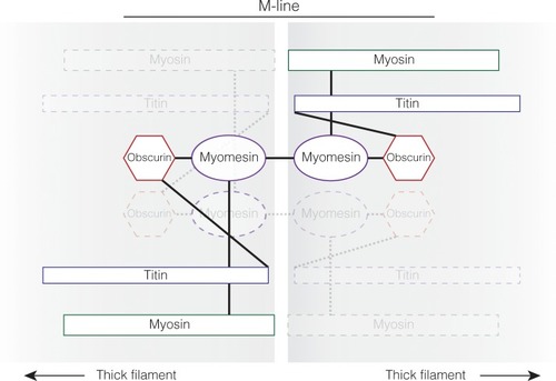
The M-line of the sarcomere diagraming the protein interactions between myosin thick filaments. The physical protein interactions through the M-line between antiparallel thick filaments are demonstrated by solid lines for one connection and faded dotted lines for the other connection between antiparallel thick filaments. Myosin and titin are incorporated into the M-line around the same time. This work suggests that myomesin is added next and requires both titin and myosin to be present for myomesin to be incorporated. Titin and myomesin together recruit obscurin, or obscurin-like 1, to the M-line [
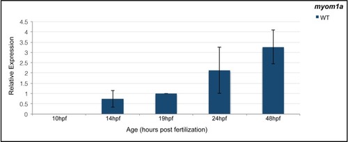
9,42].

<italic>Myomesin 1a</italic> is expressed at specific stages of myogenesis.

qPCR analysis of myom1a expression at 10, 14, 19, 24 and 48 hpf in wild-type embryos demonstrated that myom1a is not expressed until some time between 10–14 hpf (slow muscle development) and increases throughout myogenesis.

EXPRESSION / LABELING:
Gene:
Fish:
Anatomical Term:
Stage Range: Bud to Long-pec

Myomesin is incorporated into skeletal muscle around 24 hpf.

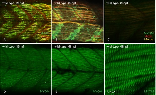
At 24 hpf, slow myosin (A) and titin (B) are incorporated and easily visible in the slow muscle fibers of wild-type embryos. Myomesin striations are observed in the parallel slow fibers of caudal somites (C). At 36 hpf, myomesin staining is seen in the developing fast fibers (D) and these striations become more organized and sharp as myogenesis continues at 48 hpf (E&F).

EXPRESSION / LABELING:
Antibodies:
Fish:
Anatomical Terms:
Stage Range: Prim-5 to Long-pec

Myomesin is organized into cardiac sarcomeres between 28 and 32hpf.

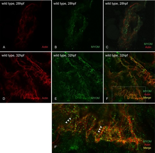
At 28hpf, we could not detect any incorporated myomesin into the sarcomeres of wild-type embryo hearts (A-C). However at 32hpf, myomesin striations are easily visualized in the hearts of wild-type embryos (D-F; white arrowheads in F’).

EXPRESSION / LABELING:
Antibody:
Fish:
Anatomical Term:
Stage Range: Prim-5 to Prim-15

Myomesin is absent in thick filament and titin mutant sarcomeres.

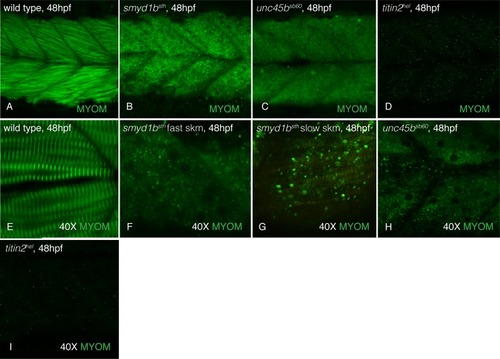
At 48hpf, myomesin is incorporated into the slow (perpendicular fibers) and fast (slanted fibers) muscle of wild-type siblings (A&E) while disorganized in smyd1btm123a (B&F), unc45bsb60 (C&H) and titin2tg287 (D&I) embryos. Increased magnification (40X) of muscle tissue demonstrates the sharp repeating striations of incorporated myomesin in wild-type siblings (E). Smyd1btm123a fast muscle (F; striations are visible in smyd1btm123a slow muscle, G), unc45bsb60 (H) and titin2tg287 (I) muscle lack myomesin striations even at 40X magnification.

Myosin and titin are required for myomesin incorporation into the sarcomere.

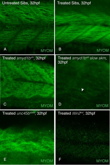
Embryos treated with tricaine to inhibit movement and contraction of skeletal sarcomeres from 18 to 32 hpf showed myomesin incorporates normally in untreated (A) and treated (B) wild-type siblings. Myomesin incorporation is absent in tricaine treated smyd1btm123a fast muscle (C; myomesin striations are visible in slow muscle (white arrowhead), D) unc45bsb60 (E), and titin2tg287 (F) mutant embryos.

Myomesin is absent in damaged skeletal muscle.

Zebrafish embryos treated with galanthamine (GAL) to induce muscle damage by hypercontraction from 28 to 48hpf demonstrated actin and myosin striations in both untreated (A&B) and treated (C&D) wild-type embryos; although GAL treated embryos exhibited fiber disorganization. Myomesin striations are visible in untreated wild-type embryos (E&F) while absent in GAL treated embryo muscle (G&H) even though actin striations, indicative of intact sarcomeres, are present (G’, white arrowheads; H’). qPCR analysis of ckmb and myom1a expression in galanthamine treated and untreated wild-type embryos demonstrated a significant decrease in the expression of both M-band genes at 48hpf (I). (Error bars are standard deviation. Expression is normalized using ef1a and wild-type at 48hpf).

<italic>Myomesin1a</italic> is significantly upregulated at early stages of myogenesis in muscle mutants.

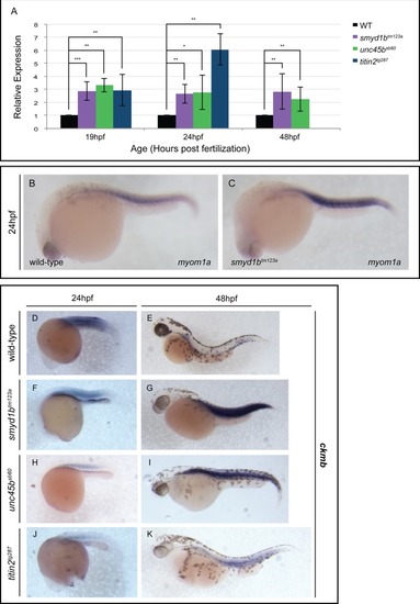
qPCR analysis at 19, 24 and 48 hpf revealed a statistically significant up-regulation of myom1a expression in smyd1btm123a, unc45bsb60 and titin2tg287 mutants relative to wild-type embryos at identical stages (A). At 24 hpf, in situ hybridization demonstrated somite-restricted myom1a expression in wild-type siblings (B), which is increased in smyd1btm123a embryos (C). At 24 hpf, wild-type (D) and smyd1btm123a (E) embryos display similar levels of muscle creatine kinase b (ckmb) expression in their skeletal muscle. Compared to wild-type embryos at 24 hpf (D), unc45bsb60 (H) and titin2tg287 (J) show a significant decrease in ckmb expression. At 48 hpf, smyd1btm123a (G), unc45bsb60 (I) and titin2tg287 (K) embryos demonstrated increased ckmb expression in their skeletal muscle when compared to wild-type siblings (E). (Error bars are standard deviation. Expression is normalized using ef1a and wild-type at corresponding stages).

Acknowledgments
This image is the copyrighted work of the attributed author or publisher, and ZFIN has permission only to display this image to its users. Additional permissions should be obtained from the applicable author or publisher of the image. Full text @ PLoS One