
FIGURE
Fig 3
- ID
- ZDB-FIG-191230-636
- Publication
- Prill et al., 2019 - Myomesin is part of an integrity pathway that responds to sarcomere damage and disease
- Other Figures
- All Figure Page
- Back to All Figure Page
Fig 3
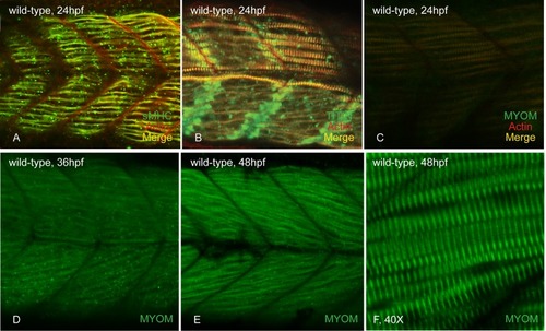
At 24 hpf, slow myosin (A) and titin (B) are incorporated and easily visible in the slow muscle fibers of wild-type embryos. Myomesin striations are observed in the parallel slow fibers of caudal somites (C). At 36 hpf, myomesin staining is seen in the developing fast fibers (D) and these striations become more organized and sharp as myogenesis continues at 48 hpf (E&F). |
Expression Data
| Antibodies: | |
|---|---|
| Fish: | |
| Anatomical Terms: | |
| Stage Range: | Prim-5 to Long-pec |
Expression Detail
Antibody Labeling
Phenotype Data
Phenotype Detail
Acknowledgments
This image is the copyrighted work of the attributed author or publisher, and
ZFIN has permission only to display this image to its users.
Additional permissions should be obtained from the applicable author or publisher of the image.
Full text @ PLoS One