
- Title
-
Zebrafish prmt5 arginine methyltransferase is essential for germ cell development
- Authors
- Zhu, J., Zhang, D., Liu, X., Yu, G., Cai, X., Xu, C., Rong, F., Ouyang, G., Wang, J., Xiao, W.
- Source
- Full text @ Development
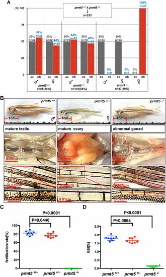
Loss of prmt5 in zebrafish reduces larval survival and generates an infertile male-like phenotype. (A) The 253 offspring of prmt5+/− (♀)×prmt5+/− (♂) matings; percentages of expected (ex, gray bars) and observed (ob, red bars). The observed percentage of prmt5−/− zebrafish (19%) was less than expected (25%). (B) The testes/ovaries and breeding tubercle in prmt5-null zebrafish (prmt5−/−) and their wild-type siblings (prmt5+/+) at 150 dpf. All prmt5−/− adults had atrophied testes, whereas prmt5+/+ adults had normal testes/ovaries. Both adult male prmt5+/+ and prmt5−/− zebrafish had breeding tubercles (arrow), which were not observed in adult female prmt5+/+ zebrafish (n=10, respectively). Red boxes indicate the positions of the magnified sections underneath. (C) The fertilization rate of prmt5+/+, prmt5+/− and prmt5−/− male zebrafish. Bars show means of eight independent experiments; error bars show s.d. (n=8, respectively). (D) The gonado-somatic indexes (GSI) of prmt5+/+, prmt5+/− and prmt5−/− male zebrafish (n=8, respectively). Differences between two groups were determined by unpaired two-tailed Student's t-test. Bars show means of eight independent experiments; error bars show s.d. PHENOTYPE:
|
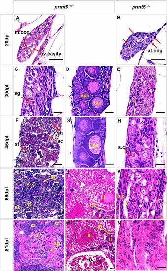
The gonads of prmt5−/− zebrafish fail to differentiate into normal testes or ovaries, as shown by histological comparisons of gonadogenesis. (A,B) At 20 dpf, prmt5−/− gonads contained a pyknotic mass and many atretic oogonia (at.oog, indicated by red arrows), while the prmt5+/+ gonads had a loosened structure with a prominent ovarian cavity (ov.cavity) and many mitotic oogonia (m.oog). (C-H) Between 30 and 45 dpf, the prmt5+/+ gonads began to differentiate into testes (C,F) or ovaries (D,G), as indicated by the presence of spermatogonia (sg), spermatocytes (sc) and spermatids (st), or by the increasing numbers of early stage IB perinucleolar oocytes(epo) and late stage IB perinucleolar oocytes (lpo). In contrast, the prmt5−/− gonads displayed a testis-like morphology containing only a few spermatogonia cysts (s.c). (I-N) Between 68 and 81 dpf, prmt5+/+ zebrafish possessed either mature testes (I,L), containing germ cells at different stages of spermatogenesis (spermatogonia, sg; spermatocytes, sc; spermatid, st; spermatozoa, sz), or mature ovaries (J,M), containing germ cells at different stages of oogenesis (IB, II, III or IV). The prmt5−/− gonads contained few spermatogonia cysts (s.c) (K) and instead contained several Sertoli cells (N). Scale bars: 20 μm; n=6 for each stage. PHENOTYPE:
|
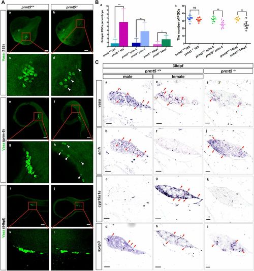
Loss of prmt5 disrupts primordial germ cell migration and causes a male-like gene expression profile during sexual differentiation. (A) Vasa in embryos at the 18-somite stage (18 s), prim-5 stage (prim5) and 54 hpf. n=80 for each stage. Arrowheads indicate the cells with relative higher gene expression. (Ba) Mean numbers of ectopic PGCs in prmt5+/+ and prmt5−/− zebrafish embryos at the 18-somite stage, prim-5 stage and 54 hpf. (Bb) Scatterplot of PGC numbers in prmt5+/+ and prmt5−/− zebrafish embryos at 18-somite stage, prim-5 stage and 54 hpf. Differences between two groups were determined by unpaired two-tailed Student's t-test. ns, not significant. *P<0.05; **P<0.01; ***P<0.001. (C) vasa, amh, cyp19a1a and sycp3 in the gonads of prmt5−/− and prmt5+/+ zebrafish during sexual differentiation (30 dpf). (a-d) Male gonads; (e-h) female gonads. Red arrowheads indicate gene expression. (b,c) Upregulation of amh and downregulation of cyp19a1a in gonads of prmt5+/+ males. (f,g) Downregulation of amh and upregulation of cyp19a1a in gonads of prmt5−/− females. (j,k) Upregulation of amh and downregulation of cyp19a1a in gonads of prmt5−/− zebrafish. (d,h) Vigorous meiosis indicated by sycp3 staining in gonads of prmt5+/+ zebrafish. (i) There are few signs of meiosis in gonads of prmt5−/− zebrafish. n=6 for each staining. Scale bars: 100 μm (A); 50 μm (C). EXPRESSION / LABELING:
PHENOTYPE:
|

Deletion of prmt5 in zebrafish leads to germ cell apoptosis. (A) Apoptotic cells in the gonads (white dashed lines) of prmt5+/+ and prmt5−/− zebrafish at 21 dpf. More apoptotic cells (green; white arrowheads) are found in the prmt5−/− zebrafish gonads than in prmt5+/+ zebrafish gonads (n=4, respectively). (B) The ratios of apoptotic cells in prmt5+/+ and prmt5−/− zebrafish at 21 dpf. (C) Apoptotic cells (arrows) in the gonads of prmt5+/+ and prmt5b−/− zebrafish at 125 pdf; n=4, respectively. (D) The ratios of apoptotic cells in prmt5+/+ and prmt5−/− zebrafish at 125 dpf. Scale bars: 20 μm. PHENOTYPE:
|
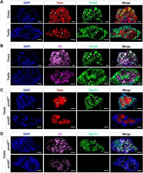
Co-expression of Prmt5 with Vasa or Zili in both ovaries and testis, and disruption of prmt5 leads to reduction of symmetric dimethylarginine levels in Vasa and Zili. (A) Vasa (red), Prmt5 (green) and DNA (DAPI staining; blue) in wild-type zebrafish ovaries (n=6) and testes (n=6) at 1 mpf. Vasa-positive and Prmt5-positive cells are indicated (white arrowheads). (B) Zili (red), Prmt5 (green) and DNA (DAPI staining; blue) in wild-type zebrafish ovaries (n=6) and testis (n=6) at 1 mpf. Zili-positive and Prmt5-possitive cells are indicated (white arrowheads). (C) Vasa (red), Sym11 (symmetric dimethylarginine antibody, green) and DNA (DAPI staining, blue) in prmt5+/+ testes (n=6) and prmt5−/− gonads (n=6) at 1 mpf. Vasa-positive and Sym11-positive cells are indicated (white arrowheads). (D) Zili (red), Sym11 (green) and DNA (DAPI staining, blue) in prmt5+/+ testes (n=6) and prmt5−/− gonad (n=6) at 1 mpf. Zili-positive and SYM11-positive cells are indicated (white arrowheads). Scale bars: 20 μm. EXPRESSION / LABELING:
PHENOTYPE:
|

The deletion of prmt5 in zebrafish results in reduction of H3R8me2s and H4R3me2s. (A,B) Vasa (red), H3R8me2s (green) (A), H4R3me2s (green) (B) and DNA (DAPI staining; blue) in prmt5+/+ testes (n=6) and prmt5−/− gonads (n=6) at 1 mpf. No H3R8me2s-positive cells (A) or no H4R3me2s-positive cells (B) were detected in prmt5−/− gonads. (C-E) Hematoxylin and Eosin staining, and H3R8me2s and H4R3me2s staining in wild-type sibling controls (prmt5+/+) and prmt5 homozygous mutants (prmt5−/−). At 90 dpf (C), the testes of prmt5+/+ zebrafish showed typical testicular morphology, with abundant spermatogonia (sg), spermatocytes (sc), spermatid (st) and spermatozoa (sz). s.t, seminiferous tube. The gonads of prmt5−/− had pyknotic cells (pc), but few spermatogonia (sg) and no spermatocytes (sc). At 104 dpf (D), in the gonads of prmt5−/− zebrafish, lumens (indicated with white dashed lines) began to enlarge, while remaining germ cells were replaced by Leydig cells (lc) and Sertoli cells (SC). The spermatogonia in the lumens gradually degenerated (Sg deg., asterisk). At 125 dpf (E), in the gonads of prmt5−/− zebrafish, lumens (indicated with white dashed lines) underwent further enlargement, germ cells were replaced by Leydig cells (lc) and the spermatogonia in the lumens degenerated. Spermatogonia, red arrows; spermatocytes, green arrows; Leydig cells, green arrowheads; Sertoli cells, red arrowheads. Scale bars: 20 μm. EXPRESSION / LABELING:
PHENOTYPE:
|

Characteristics of prmt5ihb1995/ihb1995 (M2) (5mpf) and prmt5ihb1994/ihb1995 (45dpf). (A) Appearance of body length, gonad and breeding tubercle in prmt5ihb1995/ihb1995 (M2) (5mpf). (B) Appearance of body length, gonad and breeding tubercle in prmt5+/+ and prmt5ihb1994/ihb1995 (45dpf). (C) Expression levels of vasa, dnd, ziwi, sycp3, cyp11c1, sox9a, amh and cyp19a1a in the gonads of prmt5ihb1994/ihb1995 zebrafish (45dpf) and the testes of prmt5 +/+ zebrafish (45dpf) EXPRESSION / LABELING:
PHENOTYPE:
|
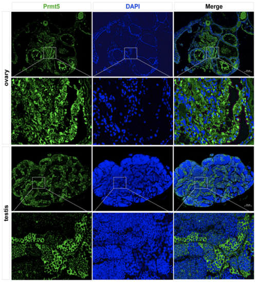
Expression of Prmt5 in testes and ovaries of adult zebrafish (4 mpf). The protein of Prmt5 was detected by Immunofluorescent staining using anti-Prmt5 antibody and the nuclei were counterstained by DAPI. |
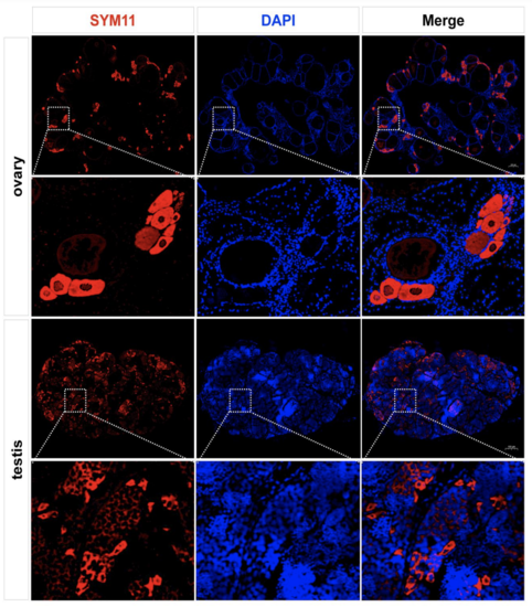
Symmetric dimethylarginine (sDMA) of proteins in testes and ovaries of adult zebrafish (4 mpf). The symmetric dimethylarginine (sDMA) of proteins were detected by immunofluorescent staining using anti-Sym11 antibody and the nuclei were counterstained by DAPI. EXPRESSION / LABELING:
|
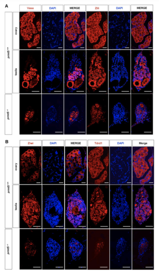
Expression patterns of Vasa, Zili, Ziwi and Tdrd1 in testes and ovaries of prmt5 +/+ and prmt5 -/- zebrafish (1 mpf). The proteins of Vasa, Zili, Ziwi and Tdrd1 were detected by immunofluorescent staining using anti-Vasa, anti-Zili, anti-Ziwi and anti-Tdrd1 antibodies, the nuclei were counterstained by DAPI. Scale bar=50 µm. |

Unillustrated author statements EXPRESSION / LABELING:
PHENOTYPE:
|