FIGURE SUMMARY
Title

Novel truncation mutations in MYRF cause autosomal dominant high hyperopia mapped to 11p12-q13.3

Authors
Xiao, X., Sun, W., Ouyang, J., Li, S., Jia, X., Tan, Z., Hejtmancik, J.F., Zhang, Q.
Source
Full text @ Hum. Genet.

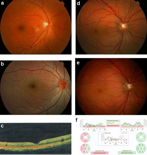
Fundus changes associated with high hyperopia in family ZOC710536.
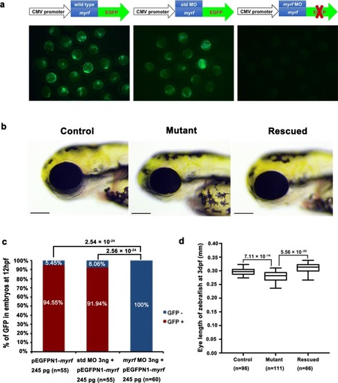
 a Normal fundus of an unrelated normal control. b Fundus photograph of V:1. The right eye for the affected individual V:1 in Fig. 1 at 6 years old with refraction of + 12D/OS and + 12D/OS, and an axial length of 17.49 mm/OD and 17.53 mm/OS. Fundus change is typical for high hyperopia with a relatively normal fovea. c OCT scan of V:1. The right eye for the affected individual V:1 in Fig. 1 at 6 years old with refraction of + 12D/OS and + 12D/OS and an axial length of 17.49 mm/OD and 17.53 mm/OS. d Fundus photos of IV: 7 at 29 years old. The right eye for the affected individual IV:7 in Fig. 1 at 29 years old, with refraction of + 10.00 DS/OD and + 10.00 DS/OS and an axial length of 17.6 mm/OD and 17.47 mm/OS. e Fundus photos of IV: 7 at 38 years old. The right eye for the individual IV:7 in Fig. 1 at 38 years old, 2 weeks after the onset of angle-closure glaucoma in the right eye (with IOP 43 mmHg before treatment). An enlarged optic disc was observed compared with the fundus photograph in D. f Heidelberg retina tomograph (HRT) results for the affected individual IV:7 from Fig. 1. The HRT result in F demonstrated partial loss of the retinal ganglion cell layer in the temporal half of the retina in the right eye. OD right eye, OS left eye

Phenotype of myrf knockdown zebrafish. a The efficiency of myrf MO knockdown of the myrf gene. Strong GFP signals were present in the left and middle images, which represent injection with myrf-pEGFPN1 plasmid alone and coinjected of myrf-pEGFPN1 plasmid with std MO, respectively. No GFP signal was detected in the embryos coinjected with myrf-pEGFPN1 plasmid and myrf MO, as shown in the right image. b Embryos with myrf knockdown showed a phenotype of small eye size (middle image) compared to the std MO-injected embryos (left image). This phenotype could be rescued by coinjection of myrf mRNA (right image). Control is larvae injected with 3 ng of std MO at 72 hpf. Mutant is larvae injected 3 ng of myrf MO at 72 hpf. Rescue is larvae coinjected with 3 ng of myrf mRNA and 106 pg of myrf MO at 72 hpf. c Proportions of embryos with GFP+ or GFP treated with 245 pg pEGFPN1-myrf, co-injection of 3 ng std MO with 245 pg of pEGFPN1-myrf and coinjection of 3 ng myrf MO with 245 pg of pEGFPN1-myrf. The p values by the Chi-squared test are shown above. d Box plot of the median and deviations of eye size in three groups. The p values by one-way ANOVA are shown above

PHENOTYPE:
Fish:
Knockdown Reagent:
Observed In:
Stage: Protruding-mouth

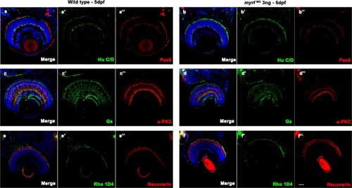
Immunostaining images of labels for different retinal cells in zebrafish at 5 dpf. Frozen sections from wild-type larvae (aa″, cc″, and ee″) and myrf MO-injected larvae (bb″, dd″, and ff″). Zebrafish larvae were labeled with Hu C/D (ganglion cell and amacrine cell marker, green, a′ and b′), Pax 6 (ganglion and amacrine precursor cell marker, red, a′′ and b′′), Gs (Müller cell marker, green, c′ and d′), PKCα (bipolar cell marker, red, c′′ and d′′), Rho 1D4 (long double cone outer segment marker, green, e′ and f′), recoverin (cone bipolar cell marker, red, e″ and f″), and DAPI (nuclei, blue). No differences were detected in retinal development between wild-type larvae and myrf morphants. Scale bars represent 100 μm

Acknowledgments
This image is the copyrighted work of the attributed author or publisher, and ZFIN has permission only to display this image to its users. Additional permissions should be obtained from the applicable author or publisher of the image. Full text @ Hum. Genet.