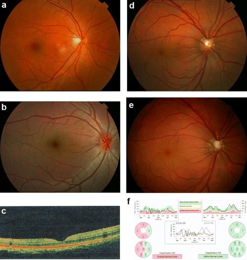
Fundus changes associated with high hyperopia in family ZOC710536. a Normal fundus of an unrelated normal control. b Fundus photograph of V:1. The right eye for the affected individual V:1 in Fig. 1 at 6 years old with refraction of + 12D/OS and + 12D/OS, and an axial length of 17.49 mm/OD and 17.53 mm/OS. Fundus change is typical for high hyperopia with a relatively normal fovea. c OCT scan of V:1. The right eye for the affected individual V:1 in Fig. 1 at 6 years old with refraction of + 12D/OS and + 12D/OS and an axial length of 17.49 mm/OD and 17.53 mm/OS. d Fundus photos of IV: 7 at 29 years old. The right eye for the affected individual IV:7 in Fig. 1 at 29 years old, with refraction of + 10.00 DS/OD and + 10.00 DS/OS and an axial length of 17.6 mm/OD and 17.47 mm/OS. e Fundus photos of IV: 7 at 38 years old. The right eye for the individual IV:7 in Fig. 1 at 38 years old, 2 weeks after the onset of angle-closure glaucoma in the right eye (with IOP 43 mmHg before treatment). An enlarged optic disc was observed compared with the fundus photograph in D. f Heidelberg retina tomograph (HRT) results for the affected individual IV:7 from Fig. 1. The HRT result in F demonstrated partial loss of the retinal ganglion cell layer in the temporal half of the retina in the right eye. OD right eye, OS left eye
|
