Image
Figure Caption

Fig. 5

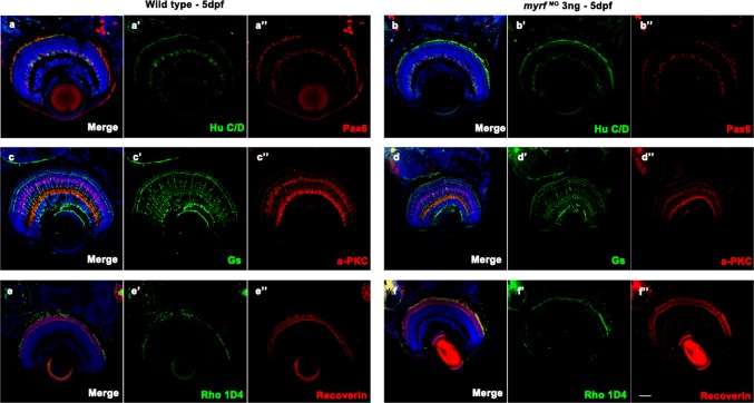
Immunostaining images of labels for different retinal cells in zebrafish at 5 dpf. Frozen sections from wild-type larvae (aa″, cc″, and ee″) and myrf MO-injected larvae (bb″, dd″, and ff″). Zebrafish larvae were labeled with Hu C/D (ganglion cell and amacrine cell marker, green, a′ and b′), Pax 6 (ganglion and amacrine precursor cell marker, red, a′′ and b′′), Gs (Müller cell marker, green, c′ and d′), PKCα (bipolar cell marker, red, c′′ and d′′), Rho 1D4 (long double cone outer segment marker, green, e′ and f′), recoverin (cone bipolar cell marker, red, e″ and f″), and DAPI (nuclei, blue). No differences were detected in retinal development between wild-type larvae and myrf morphants. Scale bars represent 100 μm

Figure Data
Acknowledgments
This image is the copyrighted work of the attributed author or publisher, and ZFIN has permission only to display this image to its users. Additional permissions should be obtained from the applicable author or publisher of the image. Full text @ Hum. Genet.