mab21l2 mutants possess complex eye defects that include small lenses, microphthalmia, coloboma, and corneal malformations. A‐L,N,O: Whole‐mount images of wild‐type (A‐E,K,L) and mab21l2 −/− (F‐J,N,O) mutant embryos. Compared with wild‐type embryos, mab21l2 −/− mutants display lens defects, microphthalmia and misshapen eyes beginning at 24 hpf (compare A‐E,K,L with F‐J,N,O). Arrowheads in I, J indicate colobomas present in mab21l2 −/− mutants. M,P,Q‐U: Ventral whole‐mount images of wild‐type (G,J) and mab21l2 −/− (H,I,K) embryos at time points indicated. Note corneal malformations and lens displacement in mab21l2 −/− mutants (arrows in P,R,S,U). Scale bars = 100 μm

mab21l2 −/− mutants possess delays in lens morphogenesis and lens growth, and do not properly express markers of mature lens cell types. A‐N: Transverse sections of wild‐type (A‐G) and mab21l2 −/− mutant (H‐N) embryos showing lens development over time. Compared with wild‐type, mab21l2 −/− mutants do not show induction of lens morphogenesis at 18SS (H); instead, comparable development is not detected until 22SS (I). Lenses remain smaller at 28 hpf (K), 36 hpf (L), 48 hpf (M), and 5 dpf (N). Note pyknotic nuclei in 28 hpf mutant (K, arrowhead). O‐Z: Whole‐mount in situ hybridizations of 48 hpf wild‐type (O‐T) and mab21l2 −/− (U‐Z) embryos. Note apparently reduced tgfβ3 (U), prox1 (X), and celf1 (Y) in mab21l2 −/− mutants, compared with wild‐type (O,R,S, respectively). Note also that mab21l2 −/− mutants lack detectable pitx3 (V), foxe3 (w), and cryaa(Z) expression. Dorsal is up in all panels. Scale bars = 50 μm

mab21l2 −/− embryos display transient increase in cell death, decreased proliferation in the developing lens. A‐C,F‐H: TUNEL stain of wild‐type (A‐C) and mab21l2 −/− mutant (F‐H) embryos. Compared with wild‐type, mab21l2 −/− mutants possess a transient increase in cell death at 22SS (F) and 26SS (G), while 48 hpf (H) embryos have equivalent proportions of dying cells to their wild‐type siblings (B, A, and C, respectively). K‐M: Quantification of TUNEL+ cells in images A‐C,F‐H. Note significant differences in proportion of TUNEL+ cells in 22SS (K; P < 0.0001) and 26SS (L; P = 0.001) but not 48 hpf (M; P = 0.63) samples. D,E,I,J: BrdU incorporation assay in wild‐type (D,E) and mab21l2 −/− mutant (I,J) embryos. Compared with wild‐type (D), mab21l2 −/− (I) mutants possess a decrease in proliferative cells in the lens at 26SS. N,O: Quantification of BrdU+ cells in images D,E,I,J. Note significant differences in the proportion of BrdU+ lens cells in 26SS (N) samples. Dorsal is up in all panels. Scale bars = 50 μm

mab21l2 −/− mutants possess colobomas of varying severity, and retain basement membrane markers in the choroid fissure. A,D: Whole‐mount images of wild‐type (A) and mab21l2 −/− mutant (D) embryos highlighting colobomas. Compared with wild‐type, mab21l2 −/− embryos show colobomas of varying severities at 5 dpf (D). B,C,E,F: Transverse (B,E) and sagittal (C,F) sections of wild‐type (B,C) and mab21l2−/− mutant (E,F) embryos at 5 dpf. Note the severe coloboma in proximal eye cup of the mab21l2 −/− mutant (E, arrow) compared with wild‐type (B). In sagittal section view, (C,F), the mab21l2 −/− retina (F) displays discontinuity of retinal lamina and failure of choroid fissure fusion (arrowhead) when compared with wild‐type (C). G‐J′: Laminin α1 localization in wild‐type (G,H′) and mab21l2 −/− (I,J′) eyes. Magenta = laminin, cyan = DAPI. Le = lens. Arrowhead marks the site of the choroid fissure. Note that wild‐type embryos (G,G′,H,H′) do not display laminin α1 at the site of the closed choroid fissure, while mab21l2 −/− mutants (I,I′,J,J′) retain laminin α1 localization at the open choroid fissure (arrowheads). Scale bars = 50 μm

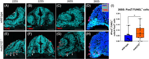
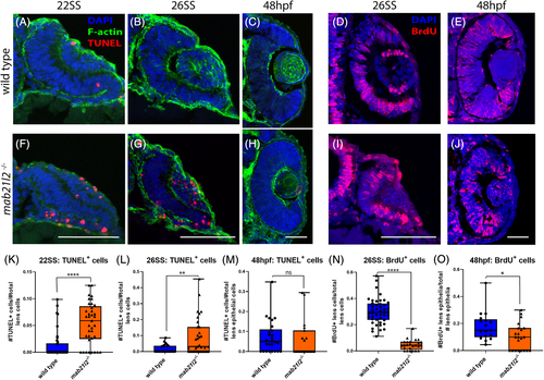
mab21l2 −/− possess elevated cell death in their optic stalk. A‐C,E‐G: TUNEL stain of wild‐type (A‐C) and mab21l2 −/− mutant (E‐G) embryos. Compared with wild‐type, 22SS mutant embryos (E,F) possess an increase in cell death in the optic stalk region of the eye. This difference persists through 26SS (G). D,H: Pax2 and TUNEL co‐stain of 26SS wild‐type (D) and mab21l2−/−(H) embryos. Compared with wild‐type (D), mab21l2 −/−embryos possess increased dying cells in the pax2+optic stalk region. I: Quantification of the proportion of pax2+ and TUNEL+ cells in D and H. Note a significantly higher (P = 0.014) proportion of pax2+ cells are TUNEL+ in mab21l2 −/− mutants when compared with wild‐type embryos. Dorsal is up in all panels. Scale bars = 50 μm
 

EXPRESSION / LABELING:
Gene:
Fish:
Anatomical Term:
Stage: 26+ somites
PHENOTYPE:
Fish:
Observed In:
Stage Range: 20-25 somites to 26+ somites

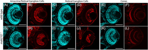
mab21l2 −/− mutants do not possess defects in retinal neuron differentiation. A‐L: Wild‐type (A‐F) and mab21l2 −/− mutant (G‐L) 3 dpf sections stained for markers of various retinal cell types. Amacrine cell/retinal ganglion cell marker HuC/D staining is similar in wild‐type (B) and mab21l2 −/− mutant (H). Retinal ganglion cell marker zn‐8 staining is similar in wild‐type (D) and mab21l2 −/− mutant (J). Cone marker zpr‐1 staining is similar in wild‐type (F) and mab21l2 −/− mutant (L). Dorsal is up in all panels. Scale bars = 100 μm
 

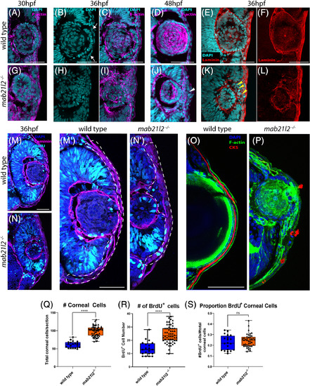
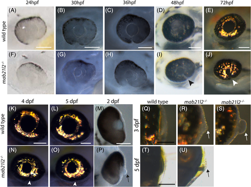
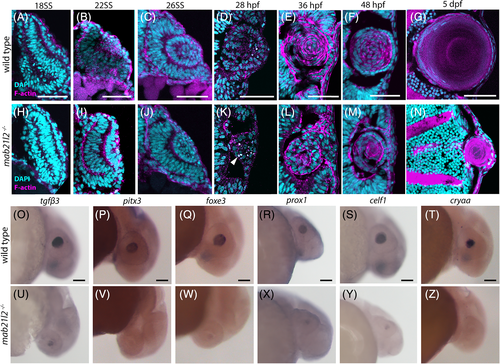
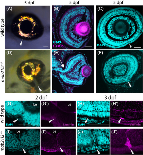
mab21l2 −/− mutants display corneal dysgenesis and failure of stromal patterning. A‐D,G‐J: Wild‐type (A‐D) and mab21l2 −/− mutant (G‐J) sections demonstrating corneal phenotypes. At 30 and 36 hpf, when compared with wild‐type eyes (A‐C), mab21l2 −/− lenses (G‐I) appear continuous with the overlying surface ectoderm. At 48 hpf, mab21l2 −/− mutants (J) possess a multilayered mass of cells (arrowhead) at the ocular surface when compared with wild‐type (D). E,F,K,L: Laminin α1 distribution in 36 hpf wild‐type (E,F) and mab21l2 −/− mutant (K,L) embryos demonstrating the presence of ectopic, nonlens, nonretinal cells in mab21l2 −/− mutants (K, arrowheads). M‐N′: BrdU incorporation assays of 36 hpf wild‐type (M) and mab21l2 −/− mutant (N) embryos. At 36 hpf, mab21l2 −/− mutants (N) possess more corneal cells, but these cells are not ectopically proliferative relative to wild‐type (M). Zooms in M′ and N′; dotted lines show area counted for quantification. O‐P: Corneal keratan sulfate (CKS) stain of 5 dpf wild‐type (O) and mab21l2 −/− mutant (P) embryos. Mutants do not properly express CKS in the stroma. Q‐S: Quantification of total and BrdU+ corneal cells in M and N. (P < 0.0001, P < 0.0001, P = 0.27, respectively). Scale bars = 50 μm

PHENOTYPE:
Fish:
Observed In:
Stage Range: Prim-15 to Long-pec
Acknowledgments
This image is the copyrighted work of the attributed author or publisher, and ZFIN has permission only to display this image to its users. Additional permissions should be obtained from the applicable author or publisher of the image. Full text @ Dev. Dyn.