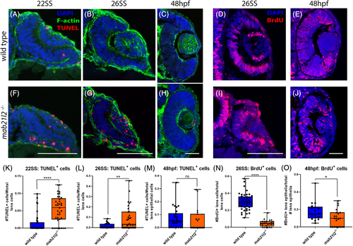
mab21l2−/− embryos display transient increase in cell death, decreased proliferation in the developing lens. A‐C,F‐H: TUNEL stain of wild‐type (A‐C) and mab21l2−/− mutant (F‐H) embryos. Compared with wild‐type, mab21l2−/− mutants possess a transient increase in cell death at 22SS (F) and 26SS (G), while 48 hpf (H) embryos have equivalent proportions of dying cells to their wild‐type siblings (B, A, and C, respectively). K‐M: Quantification of TUNEL+ cells in images A‐C,F‐H. Note significant differences in proportion of TUNEL+ cells in 22SS (K; P < 0.0001) and 26SS (L; P = 0.001) but not 48 hpf (M; P = 0.63) samples. D,E,I,J: BrdU incorporation assay in wild‐type (D,E) and mab21l2−/− mutant (I,J) embryos. Compared with wild‐type (D), mab21l2−/− (I) mutants possess a decrease in proliferative cells in the lens at 26SS. N,O: Quantification of BrdU+ cells in images D,E,I,J. Note significant differences in the proportion of BrdU+ lens cells in 26SS (N) samples. Dorsal is up in all panels. Scale bars = 50 μm
This image is the copyrighted work of the attributed author or publisher, and
ZFIN has permission only to display this image to its users.
Additional permissions should be obtained from the applicable author or publisher of the image.
Full text @ Dev. Dyn.
Your Input Welcome
Thank you for submitting comments. Your input has been emailed to ZFIN curators who may contact you if
additional information is required.
Oops. Something went wrong. Please try again later.