
- Title
-
Ioxynil and diethylstilbestrol disrupt vascular and heart development in zebrafish
- Authors
- Li, Y.F., Canário, A.V.M., Power, D.M., Campinho, M.A.
- Source
- Full text @ Environ. Int.
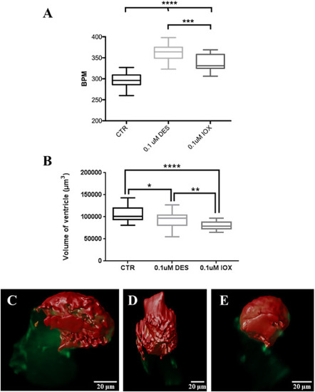
Box and whisker plot (minimum and maximum) of (A) heart beats per minute (BPM) and (B) ventricle total volume in 48 hpf zebrafish Tg(fli1:EGFP) control (CTR) and 36 h-exposure embryos to DES and IOX. Different letters indicate statistically significant difference (P < 0.05). C–E Image of the heart with the ventricle (Red) at the top and atrium (Green) at the bottom, (C) control, (D) DES treated and (E) IOX treated. One-way ANOVA was used to test for statistical significance followed by Bonferroni's multiple comparisons test *P < 0.05; **P < 0.01; ***P < 0.001; ****P < 0.0001, the same as below.
|
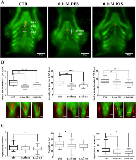
Exposure to DES and IOX affects thyroid gland and ventral aorta development. (A) Fluorescent images of the pharyngeal region of Tg(fli1:EGFP) zebrafish embryos at 72 hpf. Red and green represent, respectively, thyroglobulin and GFP immunostaining. (B) Determination of aorta diameter in three different regions as depicted in A. From left to right VA-1, VA-2 and VA-3 are represented. The lower panel below the graphs represents examples of Z-slices at each region of the aorta measured. The scale bars in the lower panel below the graphs correspond to 5 μm. (C) Measurements of thyroid follicle number, thyroid field length and the thyroid follicle area. |