
- Title
-
Pituitary adenylate cyclase-activating polypeptide (PACAP-38) plays an inhibitory role against inflammation induced by chemical damage to zebrafish hair cells
- Authors
- Kasica-Jarosz, N., Podlasz, P., Kaleczyc, J.
- Source
- Full text @ PLoS One
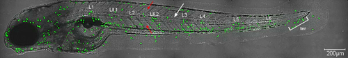
A photograph illustrating the neutrophil counting area. 5 dpf Tg(MPX:GFP) transgenic zebrafish larvae after 40 min exposure to 10 µM CuSO4 presents the area were neutrophils were quantified (green dots; in the intact larvae only single neutrophils were observed). Both neutrophils associated with investigated neuromasts (L1, LII.1, L2, LII.2, L3, L4, L5 and L6) as well as those which did not adhere directly to the neuromasts (but were sparsely found within the area encircled by the notochord [white arrow]) were counted. Dorsal and ventral myotomes marked with red arrows and terminal neuromasts (ter) were excluded from the analysis. The larvae carried myeloperoxidase promoter driving the expression of green fluorescent protein (GFP) in myeloid leukocytes (mostly neutrophils). The visualization was accomplished using a Zeiss LSM-700 confocal microscope. |
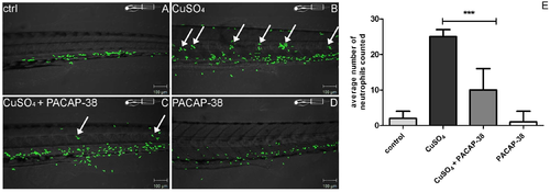
A set of microphotographs and a graph documenting inhibition of neutrophil migration towards 10 µM CuSO4 exposed hair cells resulting from co-treatment of 5 dpf Tg(MPX:GFP) zebrafish larvae (exhibiting green fluorescence in neutrophils) with 100 nM PACAP-38. (A) The control untreated larva presented normal distribution of neutrophils which were found in the ventral myotomes of the trunk and tail. (B) 10 µM CuSO4 exposure evoked the migration of the immune cells towards the midline of the body and the formation of characteristic concentrations very close to, and around, the neuromasts (arrows). (C) 100 nM PACAP-38 co-treatment resulted in the inhibition of the neutrophil migration, which was reflected by a decreased number, or complete lack of, the green fluorescent cells in the area of natural neuromast localization (arrows). (D) 100 nM PACAP-38 itself did not visibly alter the natural distribution of neutrophils. The visualization was accomplished using a Zeiss LSM-700 confocal microscope. (E) The graph presenting the influence of 100 nM PACAP-38 on the number of the neutrophils concentrated around right posterior lateral line neuromasts (PLL) (L1, LII.1, L2, LII.2, L3, L4, L5 and L6) after 10 µM CuSO4 exposure. The presented values refer to the average number of neutrophils in each group. 100 nM PACAP-38 treatment resulted in a significant, over two-fold decrease in the number of the neutrophils found singly in the area defined by notochord borders and those associated with neuromasts as compared to that determined in the 10 µM CuSO4-exposed group (one-way ANOVA, Kruskal–Wallis test with Dunn’s post-test, GraphPad Prism 5, p < 0.001). N/group = 15. |
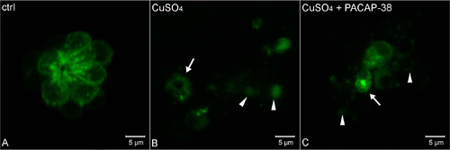
Morphology of L2 neuromast hair cells in 5 dpf Tg(pou4f3:GAP-GFP) zebrafish larvae. (A) control, (B) exposed to 10 µM CuSO4 for 40 min, and (C) exposed to the mixture of 10 µM CuSO4 and 100 nM PACAP-38 for 40 min, following 1 hour pre-incubation with 100 nM PACAP-38 only (n/group = 15). The visualization was accomplished using a Zeiss LSM-700 confocal microscope. (A) Hair cells in the control group exhibited morphology without any necrosis features. (B) Copper exposure evoked severe necrosis, resulting in hair cell rosette disintegration. Hair cells were round-shaped and swollen (arrowheads). Other groups of hair cells appeared shrunken and fragmented (arrow), suggesting the involvement of other death pathways. (C) In the PACAP-38 co-treated group, the necrosis was comparably severe. The same necrotic signs, i.e. round-shaped and swollen (arrow heads), as well as shrunken and fragmented (arrow) cells were also observed. |