
|
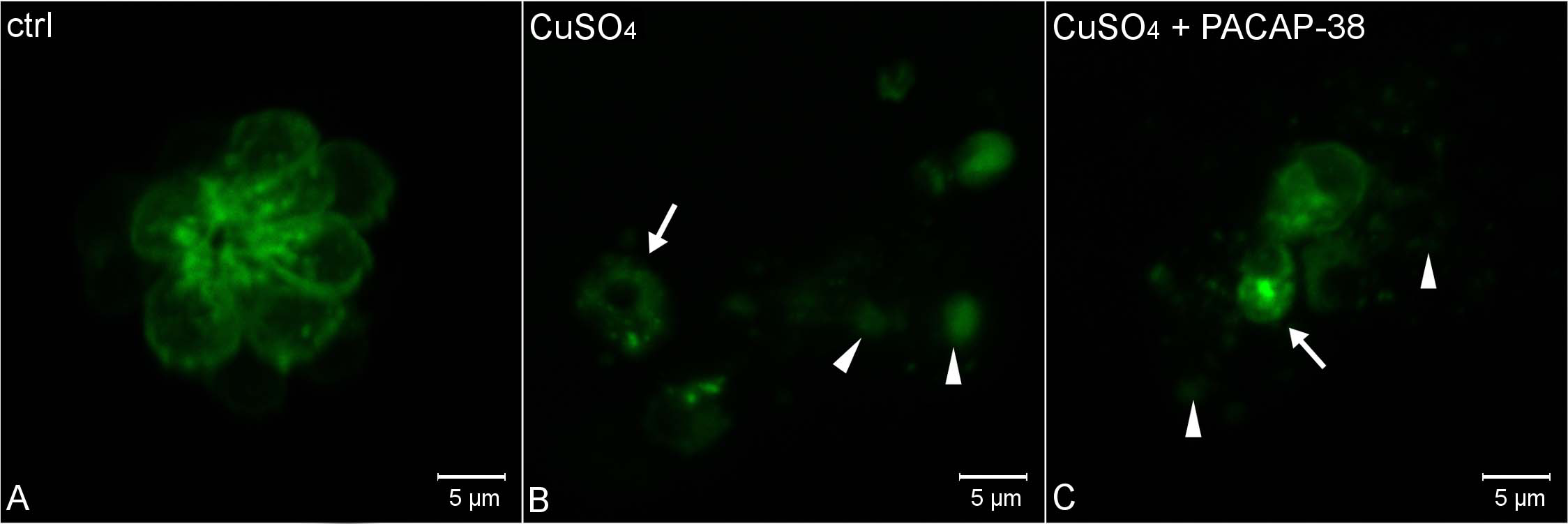
Fig. 4
Morphology of L2 neuromast hair cells in 5 dpf Tg(pou4f3:GAP-GFP) zebrafish larvae.
(A) control, (B) exposed to 10 µM CuSO4 for 40 min, and (C) exposed to the mixture of 10 µM CuSO4 and 100 nM PACAP-38 for 40 min, following 1 hour pre-incubation with 100 nM PACAP-38 only (n/group = 15). The visualization was accomplished using a Zeiss LSM-700 confocal microscope. (A) Hair cells in the control group exhibited morphology without any necrosis features. (B) Copper exposure evoked severe necrosis, resulting in hair cell rosette disintegration. Hair cells were round-shaped and swollen (arrowheads). Other groups of hair cells appeared shrunken and fragmented (arrow), suggesting the involvement of other death pathways. (C) In the PACAP-38 co-treated group, the necrosis was comparably severe. The same necrotic signs, i.e. round-shaped and swollen (arrow heads), as well as shrunken and fragmented (arrow) cells were also observed.