
- Title
-
Co-translational protein targeting facilitates centrosomal recruitment of PCNT during centrosome maturation in vertebrates
- Authors
- Sepulveda, G., Antkowiak, M., Brust-Mascher, I., Mahe, K., Ou, T., Castro, N.M., Christensen, L.N., Cheung, L., Jiang, X., Yoon, D., Huang, B., Jao, L.E.
- Source
- Full text @ Elife
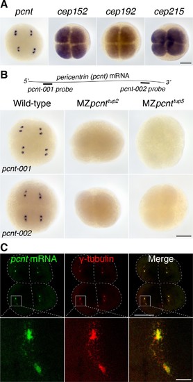
Pericentrin (pcnt) mRNA is localized to centrosomes in early zebrafish embryos. (A) RNA in situ hybridization of transcripts of different PCM components in four-cell stage zebrafish embryos. Note that while the mRNA of cep152, cep192, and cep215 displayed a pan-cellular distribution, pcnt mRNA was concentrated at two distinct foci in each cell. (B) RNA in situ hybridization showed similar dot-like patterns of pcnt transcripts with two non-overlapping antisense probes. The signals were lost in two maternal-zygotic (MZ) pcnt mutants. (C) Fluorescent RNA in situ hybridization and anti-γ-tubulin co-staining demonstrated the centrosomal localization of pcnt mRNA. n > 300 (pcnt-001 probe), n > 100 (pcnt-002 probe), n > 50 (cep152, cep192, or cep215 probe); all the embryos showed the same RNA distribution patterns as shown in the representative images. More than 100 MZpcnttup2 or MZpcnttup5 embryos were examined; none of them showed visible pcnt RNA in situ signals. Embryos were examined between 2- and 16-cell stages with representative four-cell stage embryos shown. Dashed lines delineate the cell boundaries. Scale bars: 200 µm or 25 µm (inset in C). |
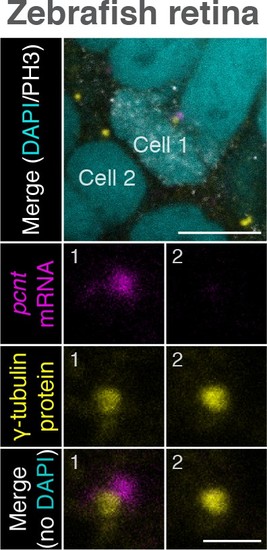
Zebrafish pcnt mRNA is localized to centrosomes of mitotic retinal neuroepithelial cells in vivo. Retinal neuroepithelial cells of 1-day-old zebrafish were subjected to pcnt FISH, anti-γ-tubulin, and anti-phospho-Histone H3 (PH3) immunostaining. Note that pcnt mRNA was localized to the centrosome of the mitotic (Cell 1, PH3+), but not of the non-mitotic cell (Cell 2). Retinas from >40 embryos were examined. Scale bars: 10 µm and 2 µm (inset). |
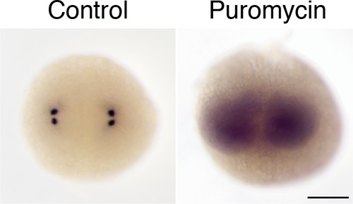
Centrosomal localization of zebrafish pcnt mRNA depends on intact polysomes. RNA in situ hybridization showed that pcnt transcripts were localized to the centrosomes in the buffer-injected embryo (Control) but were diffused throughout the cell in the embryo injected with ~1 nl of 300 µM puromycin at the one-cell stage (Puromycin). Embryos shown are at the two-cell stage. >100 embryos were examined for each condition. Scale bar: 200 µm. EXPRESSION / LABELING:
|