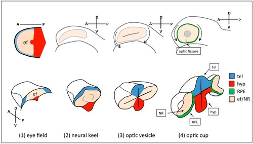
Schematic of eye morphogenesis in zebrafish. Eye field specification in the anterior portion of the neural plate (ANP) (1) is followed by the rearrangement of the tissue as the neural plate folds into a keel (2), and the evagination of the optic vesicles (3). Subsequent folding of the optic vesicle over itself leads to the formation of the optic cup (4). Top-left panel is a dorsal view with anterior to the left; the orientation of all the other panels is indicated in the figure. tel: telencephalon; hyp: hypothalamus; RPE: retinal pigment epithelium; ef: eye field; NR: neural retina.

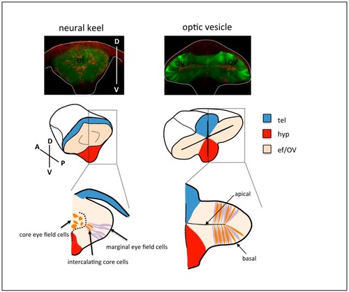
Top row: frontal views of neural keel (left) and optic vesicle (right) stage Tg{rx3::GFP} zebrafish embryos, immunostained to highlight the eye field/optic vesicles (GFP expression, green) and the apical domain of the tissue (ZO-1, red). The contour of the embryos is outlined. Middle and bottom row: schematic of cell rearrangements during optic vesicle evagination. Cells at the margin of the eye field (purple) organise as a columnar epithelium at the onset of optic vesicle evagination. Subsequent expansion of the optic vesicles is accompanied by the gradual intercalation and elongation of cells in the core of the eye field (orange) amongst marginal cells. tel: telencephalon; hyp: hypothalamus; ef: eye field; OV: optic vesicle.

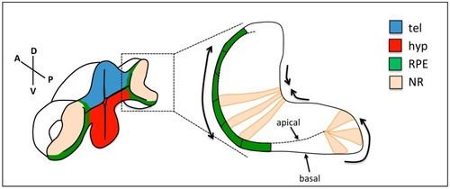
Changes in cell shape during optic cup folding. Prospective retinal pigment epithelium (RPE) cells (green) flatten, cells at the rim show apical constriction and basal lamellipodia, and neural retina (NR) cells show basal constriction. Arrows show the expected direction of tissue movement. tel: telencephalon; hyp: hypothalamus; RPE: retinal pigment epithelium; NR: neural retina.

Acknowledgments
This image is the copyrighted work of the attributed author or publisher, and ZFIN has permission only to display this image to its users. Additional permissions should be obtained from the applicable author or publisher of the image. Full text @ J Dev Biol