
FIGURE
Figure 3
- ID
- ZDB-FIG-190723-2481
- Publication
- Cavodeassi, 2018 - Dynamic Tissue Rearrangements during Vertebrate Eye Morphogenesis: Insights from Fish Models
- Other Figures
- All Figure Page
- Back to All Figure Page
Figure 3
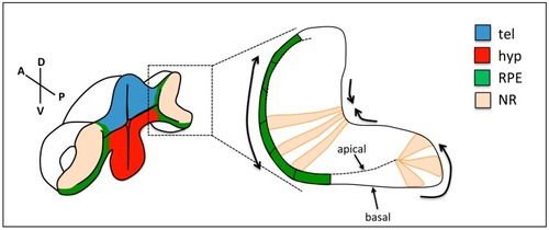
Changes in cell shape during optic cup folding. Prospective retinal pigment epithelium (RPE) cells (green) flatten, cells at the rim show apical constriction and basal lamellipodia, and neural retina (NR) cells show basal constriction. Arrows show the expected direction of tissue movement. tel: telencephalon; hyp: hypothalamus; RPE: retinal pigment epithelium; NR: neural retina. |
Expression Data
Expression Detail
Antibody Labeling
Phenotype Data
Phenotype Detail
Acknowledgments
This image is the copyrighted work of the attributed author or publisher, and
ZFIN has permission only to display this image to its users.
Additional permissions should be obtained from the applicable author or publisher of the image.
Full text @ J Dev Biol