IMAGE

Figure 2

ID
ZDB-IMAGE-190723-2515
Source
Figures for Cavodeassi, 2018
Image
Figure Caption

Figure 2

Top row: frontal views of neural keel (
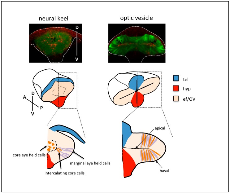
left) and optic vesicle (right) stage Tg{rx3::GFP} zebrafish embryos, immunostained to highlight the eye field/optic vesicles (GFP expression, green) and the apical domain of the tissue (ZO-1, red). The contour of the embryos is outlined. Middle and bottom row: schematic of cell rearrangements during optic vesicle evagination. Cells at the margin of the eye field (purple) organise as a columnar epithelium at the onset of optic vesicle evagination. Subsequent expansion of the optic vesicles is accompanied by the gradual intercalation and elongation of cells in the core of the eye field (orange) amongst marginal cells. tel: telencephalon; hyp: hypothalamus; ef: eye field; OV: optic vesicle.

Acknowledgments
This image is the copyrighted work of the attributed author or publisher, and ZFIN has permission only to display this image to its users. Additional permissions should be obtained from the applicable author or publisher of the image. Full text @ J Dev Biol