
- Title
-
CEP128 Localizes to the Subdistal Appendages of the Mother Centriole and Regulates TGF-β/BMP Signaling at the Primary Cilium
- Authors
- Mönnich, M., Borgeskov, L., Breslin, L., Jakobsen, L., Rogowski, M., Doganli, C., Schrøder, J.M., Mogensen, J.B., Blinkenkjær, L., Harder, L.M., Lundberg, E., Geimer, S., Christensen, S.T., Andersen, J.S., Larsen, L.A., Pedersen, L.B.
- Source
- Full text @ Cell Rep.
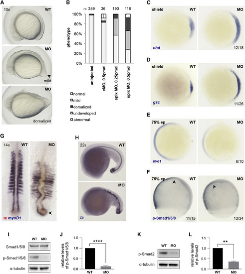
CEP128 Regulates TGF-β/BMP Signaling in Zebrafish (A) Phenotype of cep128-depleted embryos at the 10-somite stage. Embryos show mild (note the pointy tail of the embryo, arrow) to severe (egg-shaped yolk sac) dorsalized morphology. Lateral views, anterior to left. (B) Numbers for the phenotypic analysis in (A). Two to four independent experiments. (C–E) Dorsoventral patterning genes are misexpressed in cep128 splx morphants. Areas of expression of dorsal genes chd (C) and gsc (D) are increased in morphants; the ventrally expressed gene eve1 (E) is slightly decreased. Animal pole views, dorsal to right. (F) The phosphorylation level of Smad1/5/8 is slightly reduced in morphants. The thickened embryonic tissue in the morphants indicates dorsalization of the embryo (arrow). Lateral views, dorsal to right. (G) myoD1 expression (blue) shows thin and elongated somites in morphants, with adaxial cells extending around the tail bud (arrowhead); the notochord (ta expression, red) is thickened and bent. Dorsal views, anterior to top. (H) ta expression is ectopic and unstructured in the notochord and tailbud areas in cep128 morphants. (I–L) WB analysis of embryos at 75% epiboly (I and K) and quantification of relative band intensities (J and L) show that phosphorylation levels of Smad1/5/8 and Smad2 are reduced significantly in morphants compared to WT. Graphs show means ± SDs (n = 3). ∗∗p ≤ 0.01; ∗∗∗∗p ≤ 0.0001. See also Figure S3. EXPRESSION / LABELING:
PHENOTYPE:
|
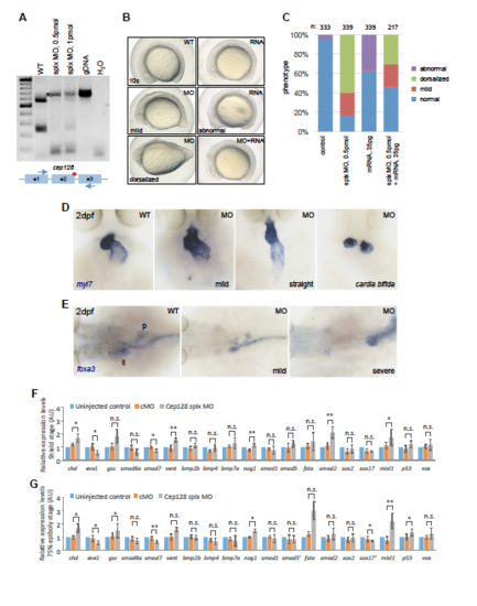
Phenotype and Rescue of cep128 Morphants. Related to Figure 2, (A) Analysis of the cep128 splx MO efficiency shows that splicing is mostly abnormal in morphants. Note the additional detected PCR product caused by alternative splicing, and inclusion of the intron (asterisk), which is also caused by the MO. Positions of the MO (red bar) and the primers (arrows) on cep128 are illustrated. (B) The dorsalized phenotype of cep128 morphants is partly rescued by injection of cep128 mRNA (right, bottom). In one third of embryos, overexpression of mRNA alone leads to an abnormal phenotype with mild to moderate defects at brain and trunk region (right, middle), which differs from the dorsalized phenotype observed with the injection of the MO (left, bottom). Left panels are taken from Figure 2A, to help with the comparison of the phenotypes. (C) Quantification of phenotypic analysis. (D) In situ hybridization with a probe against myl7 shows different defects in heart development in surviving embryos (0.25pmol MO injected) at 2 dpf, leading from a mild phenotype to few cases of cardia bifida. (E) Liver (li) and pancreas (p) development are affected by cep128 depletion (0.25 pmol) in embryos surviving to 2 dpf, as seen by foxa3 expression. (F-G) Expression levels of dorsal (gsc), ventral (eve1, vent, vox) markers and Tgf-β (smad6a, smad7, smad2, mixl1, p53) and Bmp (chd, bmp2b, bmp4, bmp7a, nog1, smad1, smad5, fsta, sox2, sox17) pathway members in cep128 splx morphants (0.5 pmol) at shield and 75% epiboly stages. * P<0.05, ** P<0.01 (one way ANOVA analysis), n>=3 independent experiments. EXPRESSION / LABELING:
|