
- Title
-
Zebrafish Larvae Model of Dilated Cardiomyopathy Induced by Terfenadine
- Authors
- Gu, G., Na, Y., Chung, H., Seok, S.H., Lee, H.Y.
- Source
- Full text @ Korean Circ J
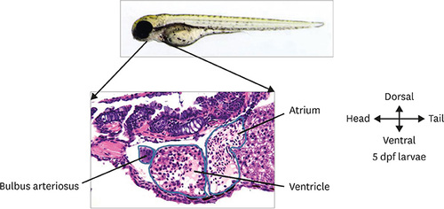
Morphology of 5 dpf zebrafish larvae after H&E staining. dpf = days post fertilization; H&E = hematoxylin and eosin. |
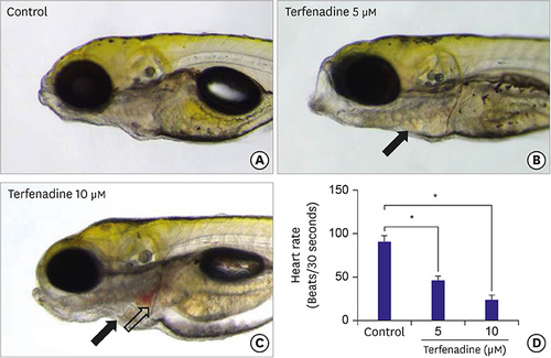
Transient terfenadine treatment reduced heart rate, inducing blood stagnation. (A) Representative image of control zebrafish larvae treated with 0.001% DMSO for 24 hours. (B, C) Representative images of zebrafish larvae treated with 5 µM (B) and 10 µM (C) terfenadine for 24 hours. Terfenadine-treated zebrafish showed enlarged heart size (black arrow) and venous congestion (hollow arrow). (D) Heart rates in control and terfenadine-treated zebrafish larvae (n=20 zebrafish/group). DMSO = dimethyl sulfoxide. *p<0.050. PHENOTYPE:
|
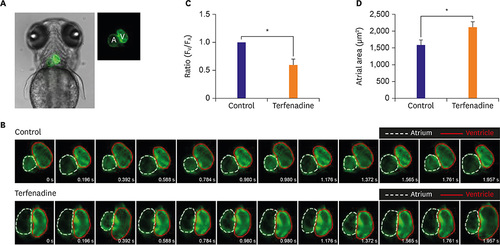
Atrioventricular dyssynchrony was induced after terfenadine treatment. (A) Representative image of cmlc-2:GFP transgenic zebrafish larvae. (B) Images taken from in vivo video recording at 4 dpf in the control (0.001% DMSO) and 24-hr terfenadine (20 μM)-treated zebrafish larvae. The interval between images in the montage is 0.196 seconds. Outlines of the atrium are shown with white dashed lines, and outlines of ventricles with red lines. (C) Arrhythmia rate of the control (0.001% DMSO) and 24-hr terfenadine (20 μM)-treated zebrafish larvae (n=20 zebrafish/group). (D) Atrial size of the control (0.001% DMSO) and 24-hr terfenadine (20 μM)-treated zebrafish larvae (n=20 zebrafish/group). A = atrium; DMSO = dimethyl sulfoxide; dpf = days post fertilization; FV/FA = fetal ventricle/fatal atrium; V = ventricle. *p<0.050. PHENOTYPE:
|
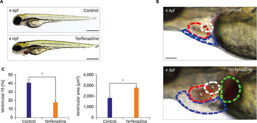
Transient terfenadine treatment impaired cardiac contraction, resulting in HF. (A) Representative images of control (0.001% DMSO) or terfenadine (20 μM)-treated zebrafish larva. (B) Lateral view of zebrafish larvae at 4 dpf. The control zebrafish (0.001% DMSO) exhibited normal cardiac morphology, whereas terfenadine (20 μM)-treated zebrafish larvae showed pericardial edema (blue circle) and venous congestion (green circle). (C) Quantification of ventricular FS in control (0.001% DMSO) and terfenadine (20 μM)-treated zebrafish larvae. Ventricle size of control (0.001% DMSO) and terfenadine (20 μM)-treated zebrafish larvae after a 24 hours treatment. n=20 zebrafish/group, scale bar=0.5 mm. a = Atrium; DMSO = dimethyl sulfoxide; dpf = days post fertilization; FS = fractional shortening; HF = heart failure; V = ventricle. *p<0.050; †p<0.010. PHENOTYPE:
|
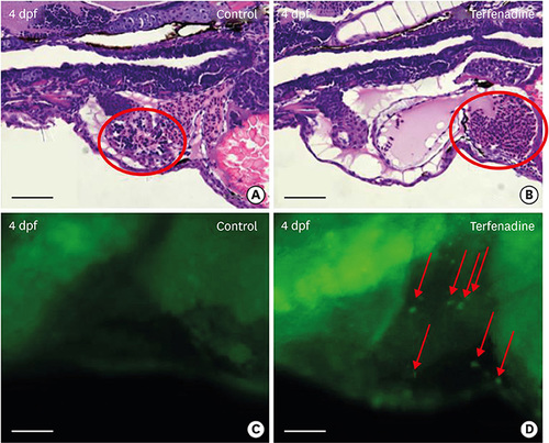
Terfenadine treatment induced apoptosis in cardiomyocytes. (A) H&E staining of longitudinal sections showed normal cardiomyocyte morphology of control (0.001% DMSO) zebrafish larvae. (B) Terfenadine (20 μM)-treated zebrafish larvae for 24 hours showed elongated and thin cardiomyocytes. (C) Detection of apoptotic cardiomyocytes by acridine orange staining in the control group. (D) Detection of apoptotic cardiomyocytes by acridine orange staining in terfenadine-treated zebrafish. Outlines of ventricles are shown with red lines. Apoptotic cells are indicated by red arrows. Scale bar=0.5 μm. DMSO = dimethyl sulfoxide; dpf = days post fertilization; H&E = hematoxylin and eosin. PHENOTYPE:
|

ZFIN is incorporating published figure images and captions as part of an ongoing project. Figures from some publications have not yet been curated, or are not available for display because of copyright restrictions. |