
|
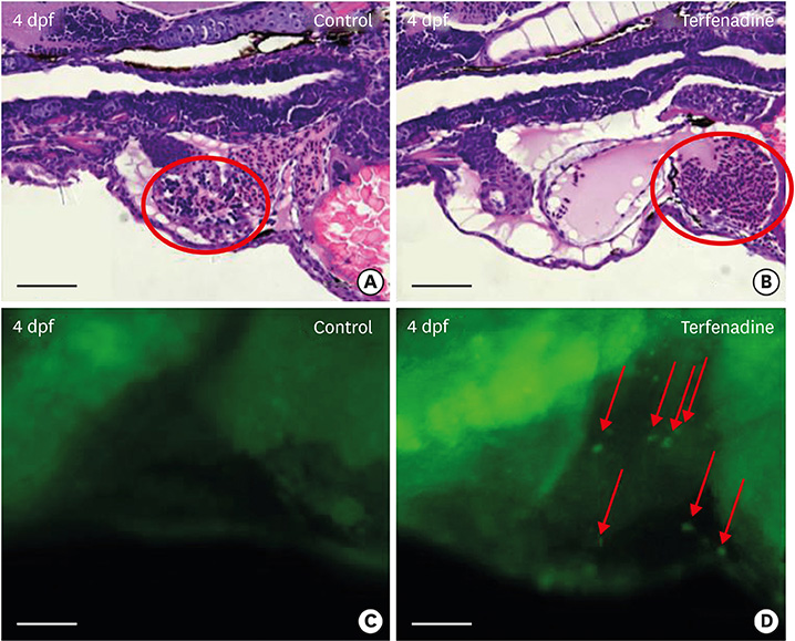
Fig. 5
Terfenadine treatment induced apoptosis in cardiomyocytes. (A) H&E staining of longitudinal sections showed normal cardiomyocyte morphology of control (0.001% DMSO) zebrafish larvae. (B) Terfenadine (20 μM)-treated zebrafish larvae for 24 hours showed elongated and thin cardiomyocytes. (C) Detection of apoptotic cardiomyocytes by acridine orange staining in the control group. (D) Detection of apoptotic cardiomyocytes by acridine orange staining in terfenadine-treated zebrafish. Outlines of ventricles are shown with red lines. Apoptotic cells are indicated by red arrows. Scale bar=0.5 μm.
DMSO = dimethyl sulfoxide; dpf = days post fertilization; H&E = hematoxylin and eosin.