
- Title
-
Signaling switch of the axon guidance receptor Robo3 during vertebrate evolution
- Authors
- Zelina, P., Blockus, H., Zagar, Y., Péres, A., Friocourt, F., Wu, Z., Rama, N., Fouquet, C., Hohenester, E., Tessier-Lavigne, M., Schweitzer, J., Roest Crollius, H., Chédotal, A.
- Source
- Full text @ Neuron

Mammalian Robo3 Does Not Bind Slits with High Affinity (A) hSlit2-D2-AP binds to COS cells expressing mammalian rRobo1A and rRobo2B but not to cells expressing mammalian Robo3A.1 or Robo3B.2. (B) Slit binding is lost in cells expressing rRobo1N88P/K90R/L130P or rRobo1L130P but restored in cells expressing mRobo3P84N/R86K/P126L or mRobo3P126L. (C) zSlit2-D2-AP binds to COS cells expressing zRobo2 or zRobo3A.1 but not to cells expressing zRobo3N83P/K85R/L125P or zRobo3L125P. (D) xSlit2-D2-AP binds to COS cells expressing xRobo3A.1 but not xRobo3N85P/K87R/L127P. (E) Scatchard analysis of Slit2 binding affinity to Robo receptors. The data shown are representative of at least three independent experiments. See also Figure S2. |

Rescue of Robo3−/− PN Neuron Midline Migration by Mammalian, but Not Nonmammalian, Robo3 (A and B) Rescue experiments by in utero electroporations of PN neurons in Wnt1::cre;Robo3lox/lox hindbrains coelectroporated at E13.5 with mouse Robo3A.1 and GFP, stained for PN marker Barhl1. Note that on the nonelectroporated side, Barhl1+ PN neurons do not migrate ventrally. By contrast, electroporated PN neurons and their axons reach the floor plate (dotted line) and/or cross it. (B) illustrates a higher magnification of the area near the floor plate. (C) E17.5 Robo3−/− hindbrain coelectroporated at E13.5 with zebrafish Robo3A.1 and GFP. None of the electroporated PN neurons or their axons leave the abberant migratory stream and/or reach the midline (dotted line). (D) Illustration of a higher magnification of the area near the floor plate. (E–H) Dorsal views of confocal z-projections of the hindbrain of 72 hpf zebrafish embryos labeled with 3A10 antibody. Anterior is toward the left. Normal midline crossing of MA axons in control (E), hsp70l:zrobo3a.1L125P (G) and hsp70l:zrobo3a.1N83PK85RL125P (H) embryos. In hsp70l:zrobo3.1 embryos, extra midline crossing events of MA axons are shown in (F). The arrows in (E) through (H) indicate normal midline crossing of MA axons; the arrowhead in (F) points to an extra MA axon midline-crossing event. Scale bars, 250 μm in (A); 50 μm in (B) and (E); 300 μm in (C); and 150 μm in (D). See also Figures S6 and S7. EXPRESSION / LABELING:
PHENOTYPE:
|

Slit2 binding on mutant Robo1 and Robo3 receptors from various vertebrate species. (A-D), hSlit2-D2-AP binds to COS cells expressing rRobo1K90R, rRobo1N88P but not mRobo3P84N or hRobo3. (E, F) chicken Slit2-D2-AP binds to COS cells expressing cRobo3A.1. (G, H) xSlit2-D2-AP binds to Xenopus Robo3N85P but does not bind to Robo3L127P. (I-L) zebrafish Slit2-D2-AP binds to COS cells expressing zRobo3A.1K85R but not zRobo3A.1N83P or zRobo3A.1N83P/K85R. (M) Scatchard analysis of hSlit2-D2-AP binding affinity to wild type and mutated Robo1 and Robo3 receptors. The data shown are representative of at least 3 independent experiments. |

Lack of high-affinity binding of hNetrin-1 to mammalian or nonmammalian Robo3 and action of a Src-family kinase and c-Abl inhibitor. after hNetrin-1 stimulation (A) Human Netrin1-AP does not detectably bind COS cells expressing either mouse, zebrafish, chick Robo3A.1 while it binds its known receptor, human DCC, with high affinity. (B) Robo3Y1019F is normally expressed at the cell surface in transfected COS cells. (C) Lckl inhibits the tyrosine phosphorylation of endogenous Robo3 induced by Netrin- 1 in P19 cells. (D) The allosteric c-Abl inhibitor GNF2 does not inhibit tyrosine phosphorylation of endogenous Robo3 after Netrin-1 stimulation in P19 cells. |

Selective rescue of Robo3-/- pontine neurons by mammalian Robo3. (A-D) E17.5 Robo3-/- hindbrain co-electroporated at E13.5 with mouse Robo3A.1 and GFP. Immunostaining for Robo3 (B) and for Barhl1/Robo3 (C). Electroporated pontine neurons send their axons (arrowheads) across the floor plate (dotted line in A-C). (C) The distance separating the floor plate from the front of migrating Barhl1 positive pontine neurons is reduced on the electroporated (rescued) side compared to the opposite (non-electroporated) side (double arrows). See text for quantification. (D) Expression levels of mRobo3A.1-V5 verified by immunoprecipitation from electroporated hindbrain protein extracts. (E-I) E17.5 Robo3-/- hindbrain co-electroporated at E13.5 with zebrafish Robo3A.1 and GFP. Immunostaining for Robo3 (F) and for Barhl1/GFP (G). Note that GFP/Robo3 positive neurons stay in the aberrant dorsal stream and do not approach the midline. (H) is a Robo3-/- embryo electroporated with zRobo3A.1 and processed as whole-mount for in situ hybridization with a zRobo3 probe. The arrows show zRobo3+ pontine neurons in the aberrant stream. (I) Expression levels of zRobo3A.1-myc (arrowhead) verified by immunoprecipitation from electroporated hindbrain protein extracts. (J, K) the mouse Robo3 lacking the CC2-CC3 domain fails to rescue ventral migration. (L, M) pontine neurons and axons from Wnt1:cre;Robo3lox/lox knockout embryo expressing Robo3Y1019F phosphorylation mutant, failed to reach the midline. Scale bars: 60μm in (M), 100 μm in (A-C), 120 μm in (E and L) and (F), 150 μm in (G), 220 μm in (J, K), 400 μm in (H) |
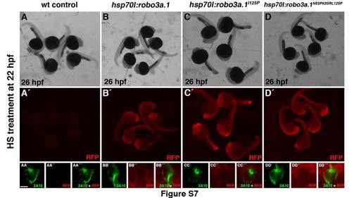
Characterization of transgenic heatshock zebrafish lines (A-D') Expression of Tdtomato as visualized by whole mount anti-RFP immunohistochemistry in control embryos and indicated transgenic lines upon heat shock treatment is shown. (AA'-DD''') High magnification of single confocal images (1 μm) is shown. Tdtomatocaax (labeled by anti-RFP antibody) is present in the cell membrane of MA neurons (labeled with 3A10 antibody). Scale bar in AA' is 5μm. |