
- Title
-
Distinct effects of inflammation on preconditioning and regeneration of the adult zebrafish heart
- Authors
- de Preux Charles, A.S., Bise, T., Baier, F., Marro, J., Jaźwińska, A.
- Source
- Full text @ Open Biol.
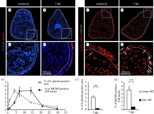
CM proliferation is correlated with the distribution of phagocytic immune cells. (a,b) Representative sections of the hearts reveal a few L-plastin-positive cells in uninjured fish in comparison to fish at 7 days post-thoracotomy (dpt) with the abundant L-plastin staining in the compact myocardium. A white dashed line separates compact (comp. MC) and trabecular (trab. MC) myocardium. (c,d) Representative sections of the heart of cmlc:DsRed2-nuc transgenic fish (red, marker of CM nuclei) labelled with the G1/S-phase marker MCM5 (green) reveal an increased mitotic activity at 7 dpt. Arrows indicate double-positive cells. (e) Quantification of the L-plastin-positive area (white circles) and of the number of proliferative CMs (black circles) in sections of uninjured hearts, and at 1, 4, 7, 14, 21 and 30 dpt. (f,g) Quantification of L-plastin-labelled leucocytes and MCM5-positive CMs in the two distinct myocardium compartments reveals a spatial correlation of both distributions at 7 dpt. (n ≥ 4 hearts; ≥2 sections per heart; ***p < 0.001). |
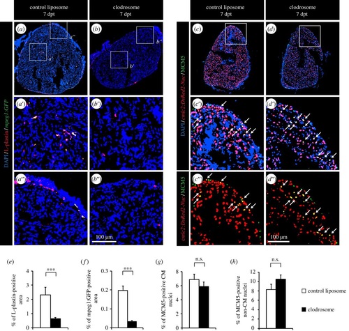
Depleting phagocytes by injection of clodronate liposomes does not affect CM proliferation. (a,b) The impact of control and clodronate liposome injection on the presence of leucocytes was tested by labelling the hearts of mpeg1:GFP transgenic fish (green) with L-plastin (red). (c,d) CM proliferation was quantified by measuring the number of CMs co-expressing DsRed2-nuc (red) and MCM5 (green). Double-positive cells are indicated with arrows. (e,f) Quantification of L-plastin- and mpeg1:GFP-positive area. (g,h) Quantification of proliferative CM and non-CM nuclei. (n ≥ 4 hearts; ≥2 sections per heart; ***p < 0.001; n.s., non-significant). |
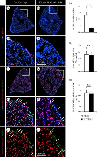
Depletion of leucocytes by PLX3397 treatment does not affect CM proliferation. (a,b) The impact of PLX3397 treatment on the presence of leucocytes was tested by labelling the hearts with L-plastin (red). (c,d) CM proliferation was quantified by measuring the number of CMs co-expressing cmlc:DsRed2-nuc (red) and MCM5 (green). Arrows indicate double-positive cells. (e) Quantification of L-plastin-positive area at 7 dpt. (f,g) Quantification of proliferative CM and non-CM nuclei at 7 dpt. (n ≥ 4 hearts; ≥2 sections per heart; ***p < 0.001; n.s. non-significant). |
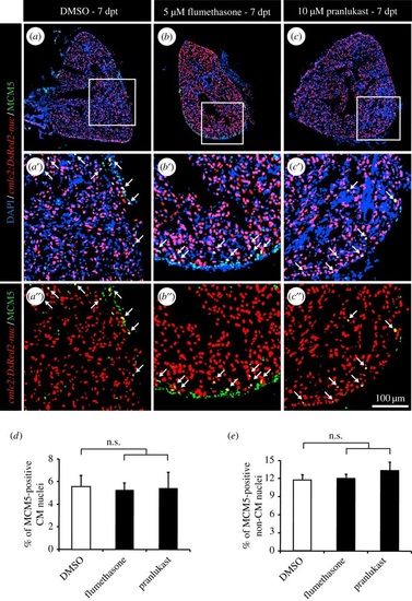
Wide anti-inflammatory drugs do not affect CM proliferation after thoracotomy. (a-c) Representative heart images of cmlc2:DsRed-nuc fish (red) treated with 0.05% DMSO (a), 5 µM flumethasone (b) or 2.5 µM pranlukast (c) labelled with the cell cycle marker MCM5 (green) at 7 dpt. Arrows indicate double-positive cells. (d,e) Quantification of MCM5-positive CM and non-CM nuclei at 7 dpt. (n ≥ 4 hearts; ≥ 2 sections per heart; n.s., non-significant). |
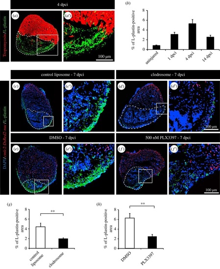
Clodronate liposome injections and PLX3397 treatment efficiently deplete phagocyte populations after cryoinjury. (a) Representative image of cryoinjured heart at 4 days post-cryoinjury (dpci) labelled with antibody against tropomyosin (red) to demarcate the remaining myocardium and L-plastin (green) to reveal leucocytes. The cryoinjured area is identified by the absence of tropomyosin (encircled by dashed line). (b) Quantification of the L-plastin-positive area in sections of uninjured hearts, and at 1, 4 and 14 dpci. (c-f) Representative images of hearts of transgenic fish cmlc2:DsRed2-nuc at 7 dpci with L-plastin staining (green). The cryoinjured area is identify by the absence of cmlc2:DsRed2-nuc expression (encircled by dashed line). (c,d) Hearts after control and clodronate liposome (clodrosome) injections. (e,f) Hearts after 0.05% DMSO or 500 nM PLX3397 treatment. (g,h) Quantification of L-plastin-positive area after clodronate liposome injection (g) and PLX3397 treatment (h) at 7 dpci. (n ≥ 4 hearts; ≥ 2 sections per heart; **p < 0.01). |
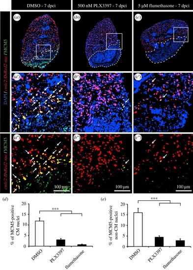
Inhibition of inflammation strongly decreases CM proliferation after cryoinjury. (a-c) Representative images of the hearts of cmlc2:DsRed-nuc fish (red) treated with 0.05% DMSO (a), 500 nM PLX3397 (b) or 5 µM flumethasone (c) labelled with the cell cycle marker MCM5 (green). The cryoinjured area is detected by the absence of cmlc2:DsRed-nuc (encircled with a dashed line). Arrows indicate double-positive cells. (d,e) Quantification of MCM5-positive CMs and non-CMs at 7 dpci. (n ≥ 4 hearts; ≥2 sections per heart; ***p < 0.001). |
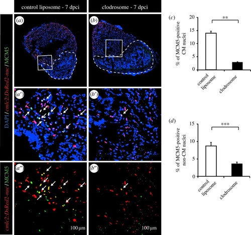
Phagocyte depletion strongly decreases CM proliferation after cryoinjury. (a,b) Representative images of the hearts of cmlc2:DsRed-nuc fish (red) injected with control liposomes (a) or injected with clodronate liposomes (b) labelled with the cell cycle marker MCM5 (green). The cryoinjured area is detected by the absence of cmlc2:DsRed-nuc (encircled with a dashed line). Arrows indicate double-positive cells. (c,d) Quantification of MCM5-positive CMs and non-CMs at 7 dpci. (n ≥ 4 hearts; ≥2 sections per heart; **p < 0.01; ***p < 0.001). |
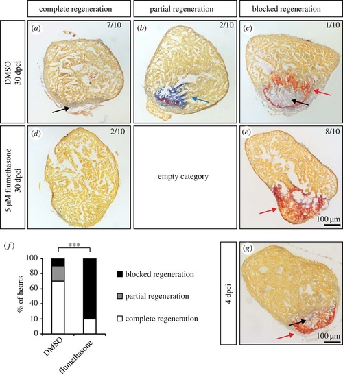
Flumethasone treatment inhibits matrix remodelling after cryoinjury and heart regeneration. (a-e) Representative sections of cryoinjured hearts at 30 dpci labelled with AFOG staining, which detects collagen in blue (blue arrow), fibrin-like matrix in red (red arrows) and muscle in orange, after 30 days of treatment with 0.05% DMSO and 5 µM flumethasone. The newly formed myocardium is indicated by black arrows. (f) Quantification of heart regeneration according to the criteria described in Materials and methods. (g) Representative example of a heart at 4 dpci labelled with AFOG staining. The cryoinjured zone in the heart at 4 dpci contains a provisional fibrotic matrix (red), which displays a similarity to the matrix of flumethasone-treated hearts at 30 dpci. (n ≥ 10; ***p < 0.001, Fisher′s exact test). |
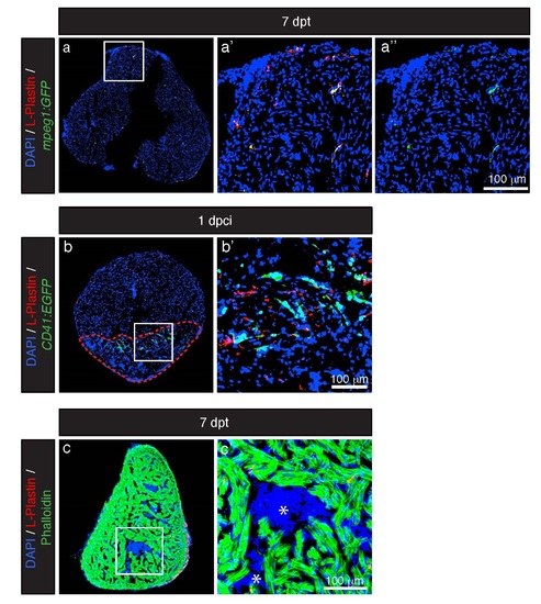
L-Plastin staining colocalizes with the macrophage marker mpeg1. (a) Representative section of the heart of mpeg1:GFP fish (green, marcrophage reporter line) at 7 dpt labeled with antibodies against L-Plastin (red, leukocyte protein) display an overlap of both markers. (b) Representative section of the heart of CD41:GFP fish (green, thrombocyte reporter line) at 1 dpci labeled with L-Plastin (red) display no overlap between both markers. Cryo-injured area is encircled with the red dashed line. (c) No L-Plastin (red) was detected in luminal blood (asterisk) of the heart at 7 dpt. |