
Fig. 1
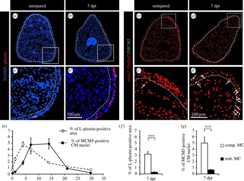
CM proliferation is correlated with the distribution of phagocytic immune cells. (a,b) Representative sections of the hearts reveal a few L-plastin-positive cells in uninjured fish in comparison to fish at 7 days post-thoracotomy (dpt) with the abundant L-plastin staining in the compact myocardium. A white dashed line separates compact (comp. MC) and trabecular (trab. MC) myocardium. (c,d) Representative sections of the heart of cmlc:DsRed2-nuc transgenic fish (red, marker of CM nuclei) labelled with the G1/S-phase marker MCM5 (green) reveal an increased mitotic activity at 7 dpt. Arrows indicate double-positive cells. (e) Quantification of the L-plastin-positive area (white circles) and of the number of proliferative CMs (black circles) in sections of uninjured hearts, and at 1, 4, 7, 14, 21 and 30 dpt. (f,g) Quantification of L-plastin-labelled leucocytes and MCM5-positive CMs in the two distinct myocardium compartments reveals a spatial correlation of both distributions at 7 dpt. (n ≥ 4 hearts; ≥2 sections per heart; ***p < 0.001). |