
Image
|
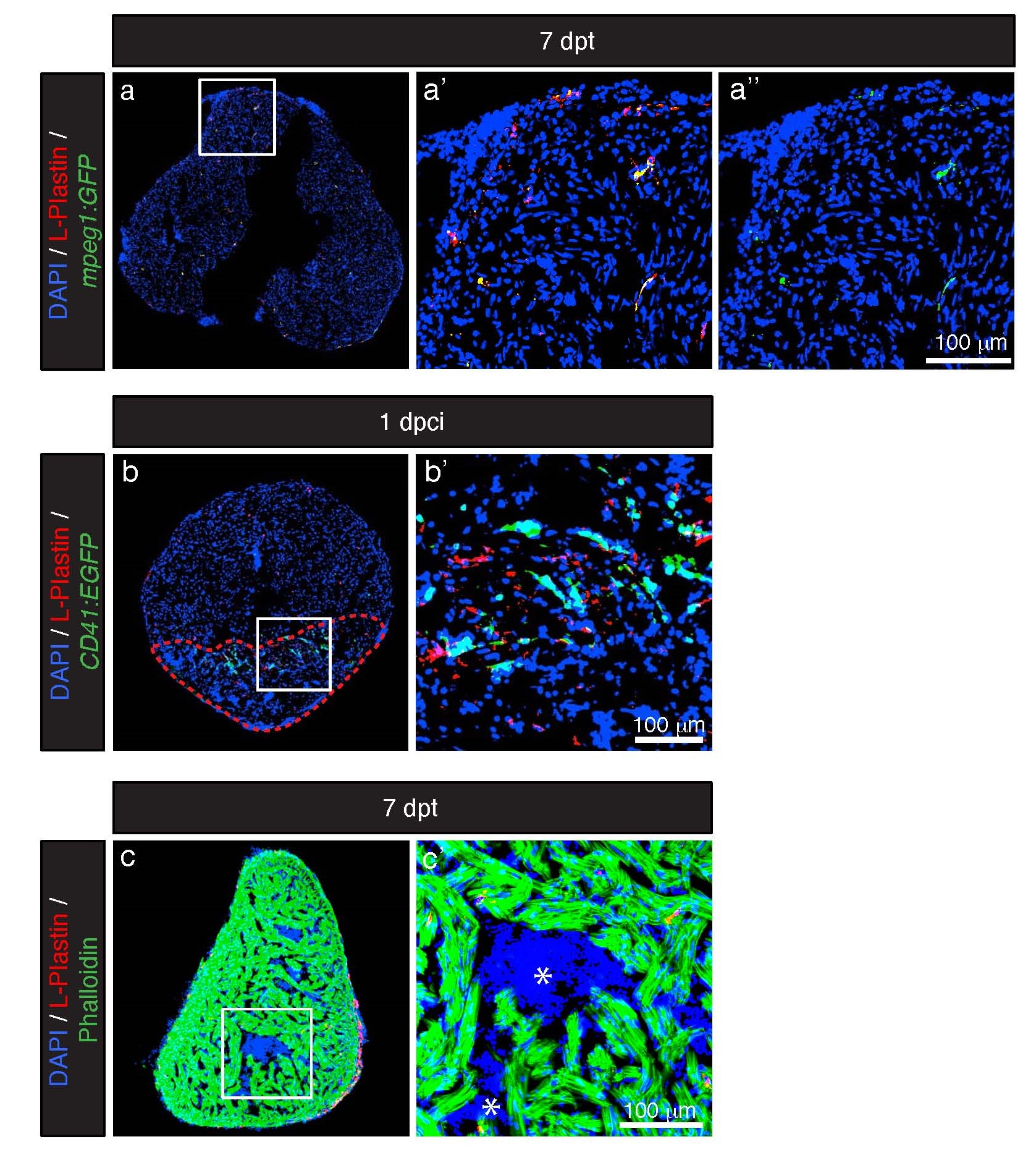
Figure Caption
Fig. S1
L-Plastin staining colocalizes with the macrophage marker mpeg1. (a) Representative section of the heart of mpeg1:GFP fish (green, marcrophage reporter line) at 7 dpt labeled with antibodies against L-Plastin (red, leukocyte protein) display an overlap of both markers. (b) Representative section of the heart of CD41:GFP fish (green, thrombocyte reporter line) at 1 dpci labeled with L-Plastin (red) display no overlap between both markers. Cryo-injured area is encircled with the red dashed line. (c) No L-Plastin (red) was detected in luminal blood (asterisk) of the heart at 7 dpt.
Acknowledgments
This image is the copyrighted work of the attributed author or publisher, and
ZFIN has permission only to display this image to its users.
Additional permissions should be obtained from the applicable author or publisher of the image.
Full text @ Open Biol.