
- Title
-
What shapes the oral jaws? Accommodation of complex dentition correlates with premaxillary but not mandibular shape
- Authors
- Hammer, C.L., Atukorala, A.D., Franz-Odendaal, T.A.
- Source
- Full text @ Mech. Dev.
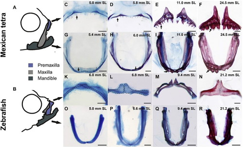
Morphological changes of oral jaws throughout growth. A-B) Schematics showing the oral jaws of the adult Mexican tetra (Astyanax mexicanus) and zebrafish (Danio rerio). The oral jaws consist of the premaxilla (blue), maxilla (light grey) and mandible (dark grey). C-R) Alizarin red and Alcian blue stained oral jaws of the Mexican tetra and zebrafish C-J) Astyanax mexicanus from 5.0 mm SL to 24.5 mm SL. Arrows indicate teeth. K-R) Danio rerio from 6.0 mm SL to 21.2 mm SL. K and L are outlined with hatched lines to help visualize the shape of the premaxilla. Scale bars are 100 µm in C-D, G-H, K, L, O-P, 200 µm in M and Q, 250 µm in E, F, I, J, N, and 500 µm in R. |
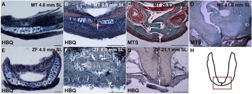
Histology of the mandibular symphysis in the Mexican tetra (MT) and zebrafish (ZF). A) A frontal section of the mandible of a 4.6 mm SL Mexican tetra. B) A transverse section of the mandible of a 9.5 mm SL Mexican tetra. C) A transverse section of the mandible of a 20.0 mm SL Mexican tetra. D) A transverse section of the mandible of an adult 61 mm SL Mexican tetra. E) A frontal section of the mandible of a 4.5 mm SL zebrafish. F) A frontal section of the mandible of an 8.0 mm SL zebrafish. G) A transverse section of the mandible of a 21.1 mm SL zebrafish. H) A schematic of a whole mount mandible with a box around the mandibular symphysis to indicate where the specimens were sectioned. A, B, E, F, and G are stained using Hall and Brunt′s Quadruple stain (HBQ) (blue, cartilage; red, bone; black, nuclei/connective tissue/epithelial cells). C and D are stained with Masson′s Trichrome stain (MTS) where green stain indicates bone. White arrows in B, C, F and G indicate the mandibular symphysis. Scale bars are as follows: 50 µm for A-B, F; 100 µm for G; 200 µm for C-D; and 20 µm for E. |
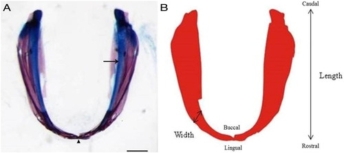
The photograph of the dissected mandible with its corresponding outline image. A) The mandible of a Mexican tetra (Astyanax mexicanus) 8.7 mm SL stained with the acid free double stain (arrow head, mandibular symphysis and arrow, Meckel′s cartilage). Scale bar is 200 µm. B) The red bitmap outline image of the mandible shown in A), which was used in the morphometrics analyses. B shows the caudal/rostral, lingual/buccal, length and width of the mandible as defined throughout the paper. |
Reprinted from Mechanisms of Development, 141, Hammer, C.L., Atukorala, A.D., Franz-Odendaal, T.A., What shapes the oral jaws? Accommodation of complex dentition correlates with premaxillary but not mandibular shape, 100-8, Copyright (2016) with permission from Elsevier. Full text @ Mech. Dev.