
Image
|
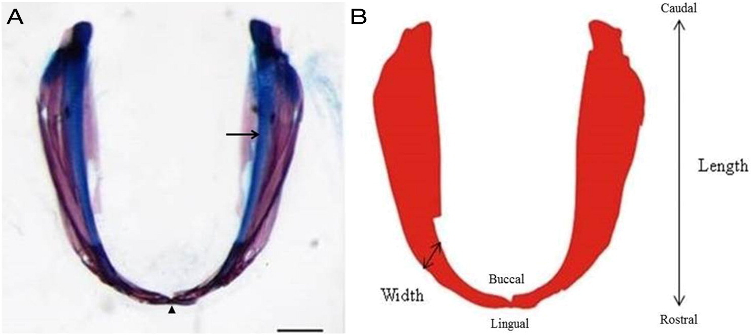
Figure Caption
Fig. 6
The photograph of the dissected mandible with its corresponding outline image. A) The mandible of a Mexican tetra (Astyanax mexicanus) 8.7 mm SL stained with the acid free double stain (arrow head, mandibular symphysis and arrow, Meckel′s cartilage). Scale bar is 200 µm. B) The red bitmap outline image of the mandible shown in A), which was used in the morphometrics analyses. B shows the caudal/rostral, lingual/buccal, length and width of the mandible as defined throughout the paper.
Acknowledgments
This image is the copyrighted work of the attributed author or publisher, and
ZFIN has permission only to display this image to its users.
Additional permissions should be obtained from the applicable author or publisher of the image.
Reprinted from Mechanisms of Development, 141, Hammer, C.L., Atukorala, A.D., Franz-Odendaal, T.A., What shapes the oral jaws? Accommodation of complex dentition correlates with premaxillary but not mandibular shape, 100-8, Copyright (2016) with permission from Elsevier. Full text @ Mech. Dev.