
Fig. 2
- ID
- ZDB-FIG-160922-20
- Publication
- Hammer et al., 2016 - What shapes the oral jaws? Accommodation of complex dentition correlates with premaxillary but not mandibular shape
- Other Figures
- All Figure Page
- Back to All Figure Page
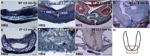
Histology of the mandibular symphysis in the Mexican tetra (MT) and zebrafish (ZF). A) A frontal section of the mandible of a 4.6 mm SL Mexican tetra. B) A transverse section of the mandible of a 9.5 mm SL Mexican tetra. C) A transverse section of the mandible of a 20.0 mm SL Mexican tetra. D) A transverse section of the mandible of an adult 61 mm SL Mexican tetra. E) A frontal section of the mandible of a 4.5 mm SL zebrafish. F) A frontal section of the mandible of an 8.0 mm SL zebrafish. G) A transverse section of the mandible of a 21.1 mm SL zebrafish. H) A schematic of a whole mount mandible with a box around the mandibular symphysis to indicate where the specimens were sectioned. A, B, E, F, and G are stained using Hall and Brunt′s Quadruple stain (HBQ) (blue, cartilage; red, bone; black, nuclei/connective tissue/epithelial cells). C and D are stained with Masson′s Trichrome stain (MTS) where green stain indicates bone. White arrows in B, C, F and G indicate the mandibular symphysis. Scale bars are as follows: 50 µm for A-B, F; 100 µm for G; 200 µm for C-D; and 20 µm for E. |
Reprinted from Mechanisms of Development, 141, Hammer, C.L., Atukorala, A.D., Franz-Odendaal, T.A., What shapes the oral jaws? Accommodation of complex dentition correlates with premaxillary but not mandibular shape, 100-8, Copyright (2016) with permission from Elsevier. Full text @ Mech. Dev.