
- Title
-
Differential Requirement for Pten Lipid and Protein Phosphatase Activity during Zebrafish Embryonic Development
- Authors
- Stumpf, M., den Hertog, J.
- Source
- Full text @ PLoS One
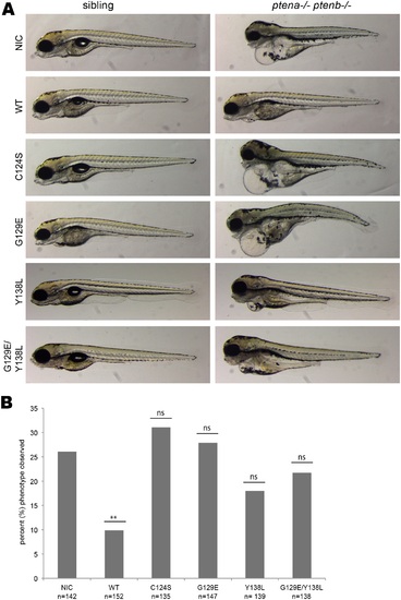
Both Pten phosphatase activities are required to rescue the ptena-/-ptenb-/- pleiotropic phenotype. Zebrafish embryos from a ptena+/- ptenb-/- incross were injected with either wild type Ptenb-mCherry, Ptenb-mCherry C124S, Ptenb-mCherry G129E or Ptenb-mCherry Y138L encoding synthetic mRNA at the one-cell stage. (A) At 4 dpf the embryos were submitted to brightfield microscopy and analyzed for the pleiotropic phenotype. Pictures show representative, genotyped embryos. Non-injected control embryos (NIC) were included for reference. (B) Quantification of the embryos showing the typical ptena-/-ptenb-/- pleiptropic phenotype at 4dpf. In the non-injected control (NIC), approximately 25% of the embryos showed the characteristic phenotype (Mendelian segregation). Only wild type Ptenb-mCherry rescued the pleiotropic ptena-/-ptenb-/- phenotype significantly at 4dpf. The statistical significance of each of the conditions compared to the non-injected control was determined using two-tailed Fisher’s exact test and is indicated in the bar graph (ns = not significant, * = p-value < 0,05, ** = p-value < 0,01, *** = p-value < 0,001). |
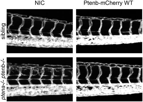
The hyperbranching vasculature phenotype, observed in Pten double homozygous zebrafish embryos at 3dpf, can be rescued by wild type Pten. Zebrafish embryos from a Tg(kdrl:eGFP) ptena+/-ptenb-/- incross were microinjected at the one-cell stage with 300 pg synthetic mRNA encoding Ptenb-mCherry. At 3dpf the embryos were analyzed for the hyperbranching vessel phenotype by confocal live imaging on a Leica TCS-SPE microscope (anterior to the left, 20x objective, 2µm z-stacks). Subsequently, the embryos were genotyped. Pictures show the trunk region distal from the urogenital opening of representative embryos; non-injected control embryos (control) were included for reference. |
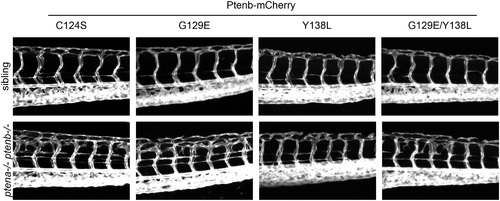
Pten lipid phosphatase activity is required to rescue the hyperbranching vasculature phenotype observed in Pten double homozygous zebrafish embryos at 3dpf. Zebrafish embryos from a Tg(kdrl:eGFP) ptena+/-ptenb-/- incross were microinjected at the one-cell stage with 300 pg synthetic mRNA encoding Ptenb-mCherry WT (not shown here, see Fig 3) or with either Ptenb-mcherry C124S, Ptenb-mcherry Y138L, Ptenb-mCherry G129E or with 150 pg of each, Ptenb-eGFP G129E and Ptenb-mCherry Y138L. At 3dpf, the embryos were analyzed for the hyperbranching vessel phenotype by confocal live imaging on a Leica TCS-SPE microscope (anterior to the left, 20x objective, 2µm z-stacks). Pictures show the trunk region distal from the urogenital opening of representative genotyped embryos. Both Ptenb WT (shown in Fig 3) and Ptenb Y138L rescue the hyperbranching phenotype observed at 3dpf. |
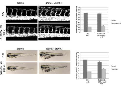
Co-injection of Ptenb-mCherry G129E and Ptenb-mCherry Y138L did not rescue developmental defects in ptena-/-ptenb-/- zebrafish embryos. Zebrafish embryos from a Tg(kdrl:eGFP) ptena+/-ptenb-/- incross were microinjected at the one-cell stage with mRNA encoding Ptenb-mCherry G129E and Ptenb-mCherry Y138L (300 pg each). At 3dpf the embryos were analyzed for the hyperbranching vessel phenotype by confocal live imaging on a Leica TCS-SPE microscope (top left panels; anterior to the left, 20x objective, 2µm z-stacks). Subsequently, the embryos were genotyped. Pictures show the trunk region distal from the urogenital opening of representative embryos; non-injected control embryos (NIC) were included for reference. Quantification of the number of embryos showing the typical ptena-/-ptenb-/- hyperbranching intersegmental vessel phenotype at 3dpf. Two-tailed Fisher’s Exact test indicated no statistically significant difference between NIC and G129E/Y138L-injected embryos (top right panel). The morphology of NIC embryos and G129E/Y138L-injected embryos was assessed at 4 dpf by brightfield microscopy (bottom left panels). Pictures show representative, genotyped embryos. Non-injected control embryos (NIC) were included for reference. Quantification of the embryos showing the typical ptena-/-ptenb-/- pleiptropic phenotype at 4dpf (bottom right panel). Two-tailed Fisher’s Exact test indicated no statistically significant difference between NIC and G129E/Y138L-injected embryos. |