
- Title
-
Mindbomb 2 is dispensable for embryonic development and Notch signalling in zebrafish
- Authors
- Mikami, S., Nakaura, M., Kawahara, A., Mizoguchi, T., Itoh, M.
- Source
- Full text @ Biol. Open
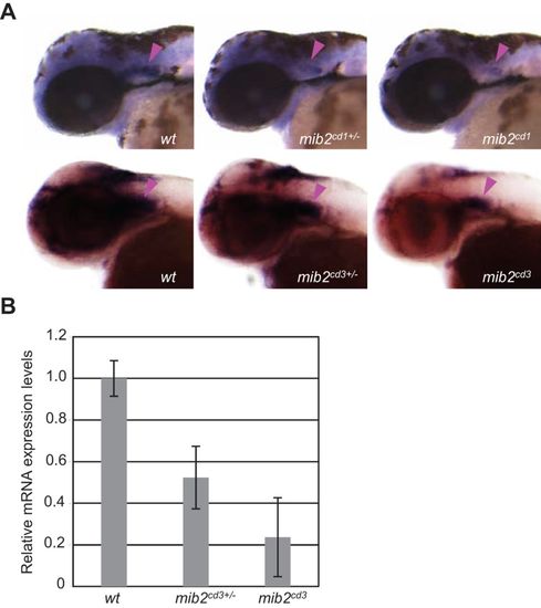
Expression of mib2 is reduced in mib2 mutant embryos. (A) Whole-mount in situ hybridization using the mib2 antisense probe in embryos at 48hpf. Arrowheads indicate mib2 expression in the ear. Head region, side views of embryos at 48hpf with anterior to the left. (B) Relative expression level of mib2 mRNA measured by q-PCR in the 48hpf-embryos. Error bars represent the mean±s.d. of three independent experiments. |
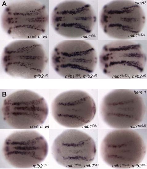
Mib2 deficiency does not significantly affect early neurogenesis and Notch signalling. Whole-mount in situ hybridization using elavl3 (A) and her4.1 (B) antisense probes at the 3 somite stage, showing no difference in neurogenesis phenotypes between wild-type and mib2cd3 embryos or between the mib1 mutants and mib1/mib2 double mutants. All panels show top views of embryos with the anterior to the left. EXPRESSION / LABELING:
PHENOTYPE:
|
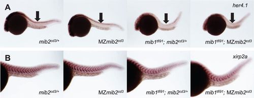
Maternal Mib2 does not compensate for the zygotic loss of Mib2. Whole-mount in situ hybridization using her4.1 (A) and xirp2a (B) antisense probe at 24hpf. Maternal deletion of mib2 does not affect expression of her4.1 or xipr2a. Whole embryo (A) and trunk region (B) are shown. Arrows show expression of her4 in the trunk neural tube. Side views of embryos with anterior to the left. |
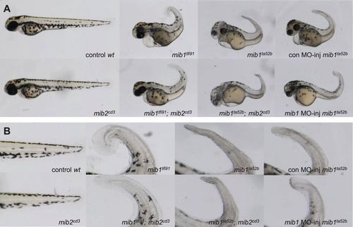
Mib2 neither regulates melanophore development nor antagonizes Mib1ta52b protein. Whole embryos (A) or enlarged views (B) of tail region in A. mib2 deletion did not enhance pigmentation loss in mib1tfi91, nor did it recover pigmentation in mib1ta52b mutants. All panels show side views of embryos with anterior to the left. PHENOTYPE:
|
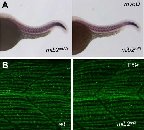
Muscle structure is normal in mib2 mutant embryos. (A) Whole-mount in situ hybridization using the myoD in trunk region. (B) Slow muscle fibre myosin in trunk muscles as revealed by F59 antibody. mib2 mutant embryos did not show any changes in the pattern or level of their staining. All panels show side views of embryos with anterior to the left at 24hpf. |