
- Title
-
Finite element modelling predicts changes in joint shape and cell behaviour due to loss of muscle strain in jaw development
- Authors
- Brunt, L.H., Norton, J.L., Bright, J.A., Rayfield, E.J., Hammond, C.L.
- Source
- Full text @ J. Biomech.
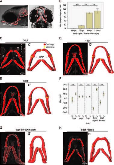
Changes to jaw joint morphology between 3 and 5 dpf and the effect of muscle activity on joint shape. (A): Brightfield lateral and ventral view of 5 dpf zebrafish expressing Tg(Col2a1aBAC:mcherry) cartilage marker MC=Meckel′s cartilage, PQ=palatoquadrate, CH=ceratohyal. (B): The number of mouth openings per minute at 58 h post-fertilisation (hpf), (n=10), 72 hpf (n=6), 96 hpf (n=6) and 120 hpf (n=6). ns= not significant, ***=Pd0.001. (C, D, E): Confocal image stacks of ventral zebrafish jaws at 3 (C), 4 (D), and 5 dpf (E), marked with a Tg(Col2a1aBAC:mcherry) cartilage marker. The medial and lateral sides of the elements are labelled M and L and the anterior and posterior surfaces of the elements are labelled A and P. IZ marks the interzone. (C′, D′, E′): 3D Avizo reconstructions from the confocal datasets: cartilage marked red, interzone marked yellow. RAP denotes the retroarticular process of the Meckel′s cartilage (F): Box and whisker plot showing the interval between the anterior MC and posterior PQ elements on the medial and lateral side of the joints at 3 dpf (n=12), 4 dpf (n=16), 5 dpf (n=13) and in 5 dpf myod (n=4) and 5 dpf anaesthetised zebrafish (n=8), ns=not significant, ***=Pd0.001. Negative measurements indicate an overlap of the anterior MC and posterior PQ elements. (G, H): confocal image stacks of the ventral jaws of 5 dpf myod mutant (G) and 5 dpf anaesthetised zebrafish (H) marked by Tg(Col2a1aBAC:mcherry) cartilage marker. (G′, H′) 3D Avizo reconstructions from the confocal datasets. (For interpretation of the references to color in this figure legend, the reader is referred to the web version of this article.) |
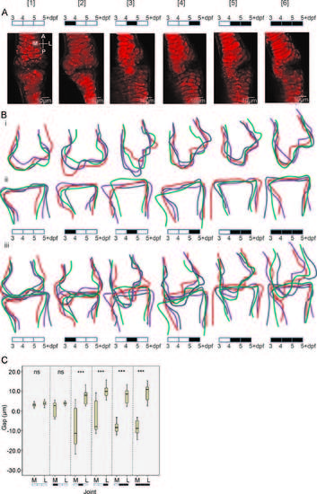
Imaging and quantification of joint shape. (A); zebrafish 5+ dpf jaw joints labelled with the Tg(Col2a1aBAC:mcherry) cartilage marker, anaesthetised with MS222 for varying time periods between 3 and 4 [2], 4 and 5 [3], 5 and 5+ [4], 4–5+ [5] and 3–5+dpf [6]. (5+dpf=128 hpf). A black box on the 3–5+dpf timeline indicates anaesthetisation by MS222 and an empty box indicates no MS222 treatment. The medial and lateral side of the elements are labelled M and L and the anterior and posterior surfaces of the elements are labelled A and P. (B): Outline of the 5+ dpf jaw joint shape after each anaesthetic treatment, anterior Meckel′s cartilage joint element (Bi), posterior Palatoquadrate joint element (Bii), and the extent of the joint element overlap (Biii) (n=4, each colour representing an individual fish). All outlines are to the same scale. (C): Box and whisker plot of the interval between the anterior MC and posterior PQ elements on the medial and lateral side of the joints at 5+dpf (n=13, 13, 10, 10, 16, 16, 18, 16, 8, 8, 12, 14, 8, 8), ns=not significant, ***=Pd0.001. Negative measurements indicate an overlap of the anterior MC and posterior PQ elements. |