
- Title
-
Zebrafish adult-derived hypothalamic neurospheres generate gonadotropin-releasing hormone (GnRH) neurons
- Authors
- Cortés-Campos, C., Letelier, J., Ceriani, R., Whitlock, K.E.
- Source
- Full text @ Biol. Open
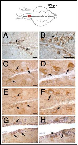
GnRH antibodies recognize cells in the anterior preoptic region of the adult brain. Brain sections (A,B, cryostat sections; C-G, paraffin sections) of different animals immuno-stained with different antibodies against GnRH: (A,B) anti-GnRH (LRH13); (C,D) anti-GnRH (Hu11B); (E,F) anti-mGnRH (Sigma-Aldrich); (G,H) anti-sGnRH (BB8). All antibodies show immunoreactivity in neurons (arrows) localized in the hypothalamic POA (diagram, red). Scale bars: 50µm. |
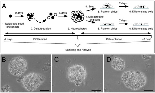
Neurospheres can be generated from the hypothalamus of adult zebrafish. (A) Neural progenitors were disaggregated (black cells), plated, and maintained in proliferation media for 7day (A1 to A3 neurospheres, 7 to 0days). Cells were then seeded (A4 to A6, upper panel) on coated chamber wells and changed to differentiation media for 7days (0 to +7days). Alternatively, neurospheres (A3) were disaggregated and seeded (A4 to A6, bottom panel) on coated chamber wells and changed to differentiation media for 7days with or without hormonal treatment (0 to +7days). (B-D) 7days in vitro bright field images of neurospheres (A3). Scale bar 30µm. |

Hypothalamic neurospheres express neural progenitor markers. Undifferentiated 7days in vitro neurospheres. Cells expressing nestin and GFAP are located principally at the center of the neurospheres, (A-C, arrows). Sox2 and vimentin, markers of undifferentiated neural progenitors, are expressed in the neurospheres (D-F, arrows). Neurospheres are highly proliferative, expressing PCNA (green) in many of the GFAP positive (red) cells (G-I, arrows). Undifferentiated neurospheres reactive to GFAP show a reduced number of neurons (positive for HUC) (J-L, arrows). Scale bar 25µm; n=3 plates derived from different cultures. |
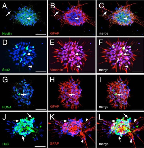
Hypothalamic progenitors generate glial and neuronal cells. (A-F) Neurospheres differentiated after 7days in vitro. (A-C) Differentiated cells surround the core of cells positive for nestin (arrow head) and extend cellular projections that are reactive to GFAP (arrows). (D-F) The reduced number of Sox2 positive cells (arrows) and the high number of cell processes reactive to Vimentin (arrow heads) suggests that progenitors have differentiated into glial cells. (G-L) Neurospheres reactive to GFAP show (G-I) a reduced number of cells positive for PCNA (arrows), (J-L) high number of neurons (arrows) and differentiated glial cells (arrow heads). Scale bar 25µm, n=3 plates derived from different cell cultures. |
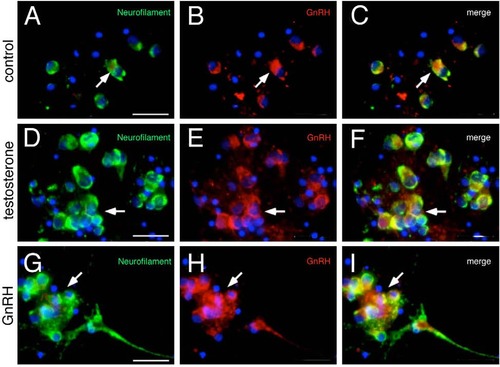
Hypothalamic progenitors can differentiate GnRH neurons. (A-C) Neurospheres differentiated for 7days in vitro without hormonal stimulus. Neurospheres generate GnRH neurons (arrows). (D-F) Differentiated 7days in vitro neurospheres, stimulated for 5days with 10µM testosterone. Testosterone increases the number of GnRH neurons in NSC cultures (arrows). (G-I) Differentiated 7days in vitro neurospheres, stimulated for 5days with 10nM GnRH increases the number of GnRH neurons in NSC culture (arrows). Scale bar 25µm. |

The hypothalamus of the adult zebrafish contains newly differentiated GnRH cells. Horizontal sections showing the anterior region of the parvocellular preoptic nucleus (PPa), with DAPI labeling (blue). (A-C) HuC labeling in a HuC:GFP background (green, arrows) and vimentin labeling (red, arrowhead) do not co-localize. The vimentin positive cells line the edges of the ventricles. (D-F) HuC (green) and Sox2 labeling (red, arrows) co-localizes in some cells (arrows) located closer to the border of the ventricle (arrows). (G-I) Section showing GnRH cell body labeling (green, box, asterisk) and labeling of processes (green, arrows), Sox2 labeling (red, box, asterisk) in cells located several cell diameters from the border of the ventricle (arrows) co-localizes with the GnRH labeling (merge). All Images: anterior is to the left. Scale bar=30µm. |
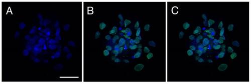
Undifferentiated secondary neurospheres do not express GnRH. Undifferentiated secondary neurospheres were labeled with DAPI, anti-GnRH antibody (LHR13; A, no expression detected), anti-Sox2 antibody (B, green), with all channels shown in C. All Images: anterior is to the left. Scale bar=30µm. |