
|
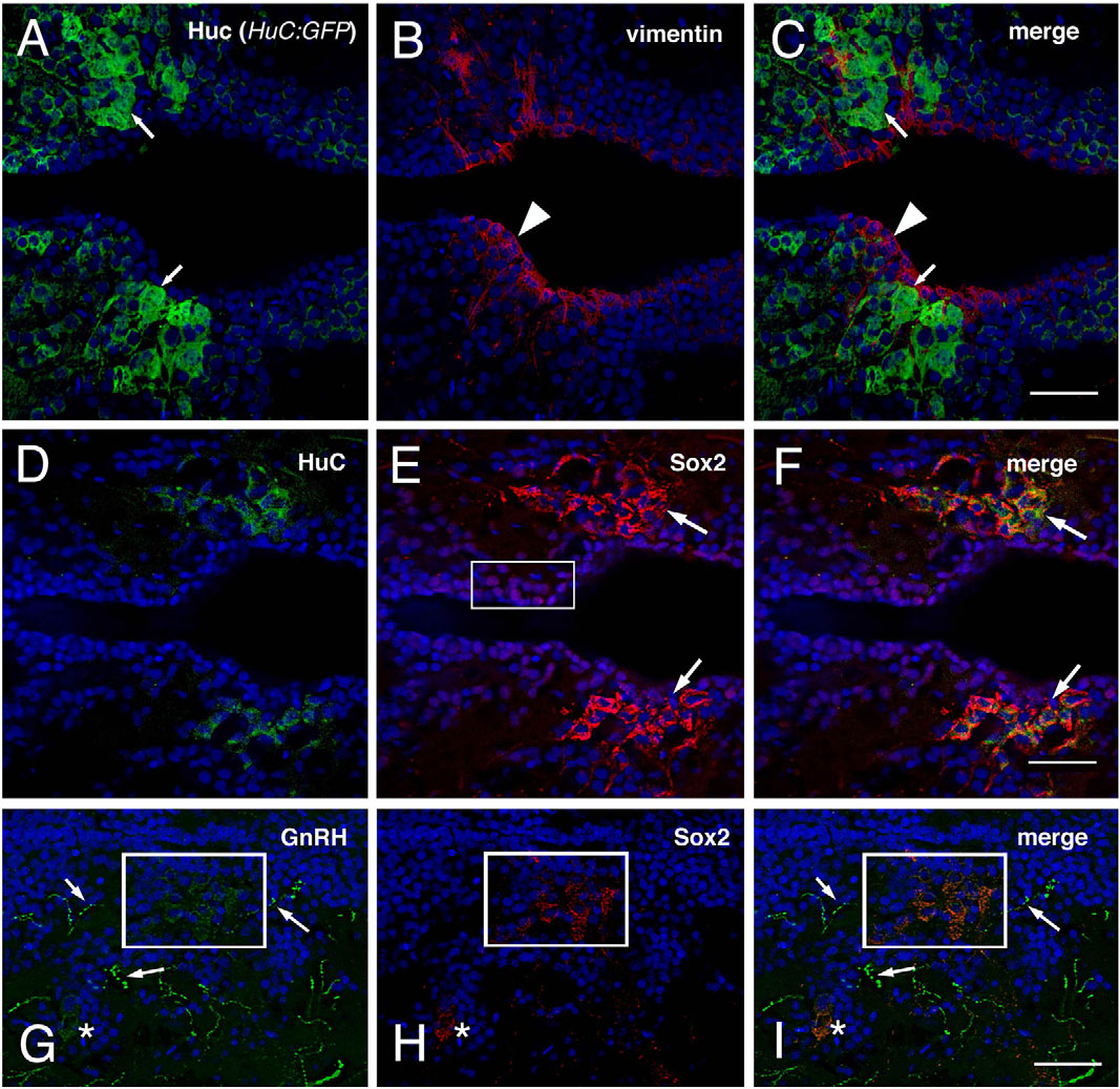
Fig. 8
The hypothalamus of the adult zebrafish contains newly differentiated GnRH cells. Horizontal sections showing the anterior region of the parvocellular preoptic nucleus (PPa), with DAPI labeling (blue). (A-C) HuC labeling in a HuC:GFP background (green, arrows) and vimentin labeling (red, arrowhead) do not co-localize. The vimentin positive cells line the edges of the ventricles. (D-F) HuC (green) and Sox2 labeling (red, arrows) co-localizes in some cells (arrows) located closer to the border of the ventricle (arrows). (G-I) Section showing GnRH cell body labeling (green, box, asterisk) and labeling of processes (green, arrows), Sox2 labeling (red, box, asterisk) in cells located several cell diameters from the border of the ventricle (arrows) co-localizes with the GnRH labeling (merge). All Images: anterior is to the left. Scale bar=30µm.