
Image
|
Figure Caption
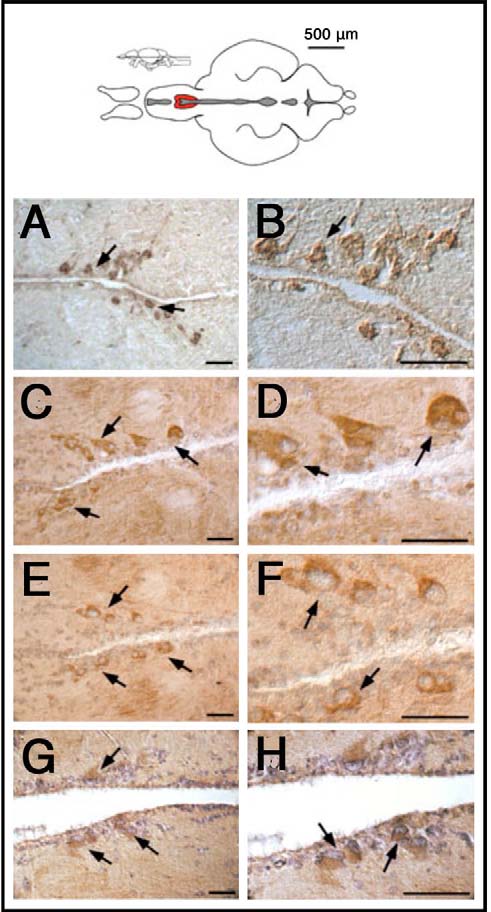
Fig. 1
GnRH antibodies recognize cells in the anterior preoptic region of the adult brain. Brain sections (A,B, cryostat sections; C-G, paraffin sections) of different animals immuno-stained with different antibodies against GnRH: (A,B) anti-GnRH (LRH13); (C,D) anti-GnRH (Hu11B); (E,F) anti-mGnRH (Sigma-Aldrich); (G,H) anti-sGnRH (BB8). All antibodies show immunoreactivity in neurons (arrows) localized in the hypothalamic POA (diagram, red). Scale bars: 50µm.
Acknowledgments
This image is the copyrighted work of the attributed author or publisher, and
ZFIN has permission only to display this image to its users.
Additional permissions should be obtained from the applicable author or publisher of the image.
Full text @ Biol. Open