
- Title
-
Conserved Noncoding Sequences Regulate lhx5 Expression in the Zebrafish Forebrain
- Authors
- Sun, L., Chen, F., Peng, G.
- Source
- Full text @ PLoS One
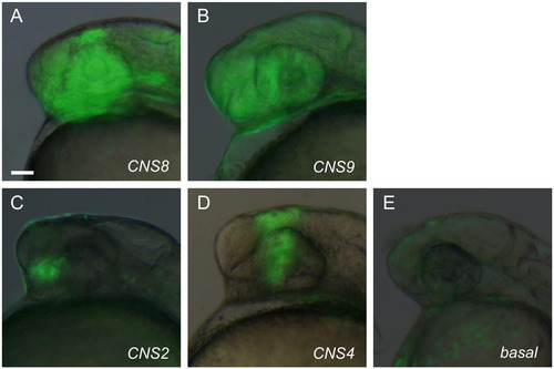
Region specific enhanver activity of the identified CNSs. (A-B) CNS8 and CNS9, located in the vicinity of the lhx5 promoter region give rise to broad reporter EGFP expression in the forebrain regions. (C) CNS2 located approximately 50 kb upstream of the lhx5 coding region gives rise to restricted EGFP signal in the anterior ventral forebrain. (D) CNS4 located 40 kb upstream of the lhx5 coding region, gives rise to restricted EGFP expression in the diencephalic region. (E) Vector construct gives rise to basal non-tissue specific EGFP expression in transient expression assay. Lateral view of the forebrain regions of embryos at 24 hpf, anterior to the left. Scale bar: 50µm. EXPRESSION / LABELING:
|
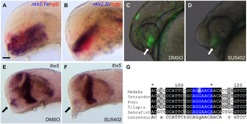
CNS2 contains hypothalamic enhancer activity and responses to FGF signaling. (A-B) Double in situ hybridization results indicate CNS2 contains hypothalamic enhancer activity. The hypothalamic marker nkx2.1a and nkx2.2b are stained in dark blue, reporter egfp stained in red. (C-D) SU5402 treatment severely reduces CNS2 activity. Vehicle DMSO treated embryos show restricted hypothalamic EGFP reporter expression (pointed by the arrow in C). Embryos treated with the FGF signaling inhibitor SU5402 during the segmentation stage (10-24hpf) show minimal EGFP signals in the hypothalamic region (arrow in D, n = 48/55). (E-F) SU5402 treatment down-regulates endogenous lhx5 expression in the hypothalamic region. Endogenous lhx5 shows robust expression in the hypothalamic region (pointed by the arrow in E). SU5402 treatment during the segmentation stage down-regulates lhx5 expression in the hypothalamic region (arrow in F, n = 25/28). (G) Multiple sequence alignments of the CNS2 region in the five teleost species. The identified FGF downstream factor Pea3 binding site is highlighted in blue. Lateral view of the forebrain regions of embryos at 24 hpf (A-F), anterior to the left. Scale bar: 40µm in A-B; 50µm in C-D. |
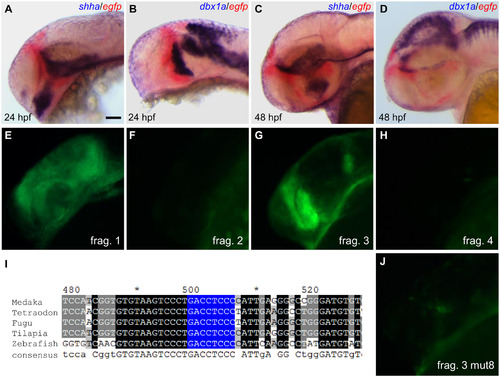
CNS4 contains pre-thalamic enhancer activity and relies on a zic binding site. (A-D) Double in situ hybridization results indicate CNS4 contains pre-thalamic enhancer activity. ZLI (zona limitans intrathalamica) marker shha and thalamic marker dbx1a are stained in dark blue, reporter egfp stained in red. CNS4 driven EGFP expression abuts anteriorly to the ZLI and thalamus in the 24hpf (A-B) and 48hpf (C-D) stage embryos. (E-H) The third fragment of the four subdivisions of the CNS4 sequence contains pre-thalamic enhancer activity. (I) Multiple sequence alignments of the CNS4 region in the five teleost species. The identified zic binding site is highlighted in blue. (J) Mutation of the 8bp zic binding site in the prethalamic enhancer abolished its activity. Lateral view of the forebrain regions of embryos at 24 hpf (A-B, E-H, and J) or 48 hpf (C-D), anterior to the left. Scale bar: 50µm. |
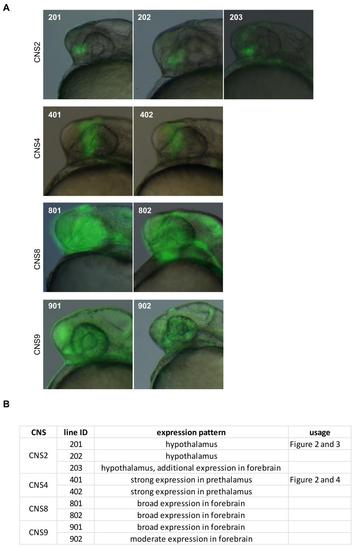
Stable transgenic lines carrying CNS elements. (A) Figure panels illustrate established transgenic lines carrying corresponding CNS elements. Line id is indicated on the upper left corner on the figure panel. Lateral view of the forebrain regions of embryos, anterior to the left. (B) Text description of the established transgenic lines. The line id used in experiments described in the Result section is indicated. |