
|
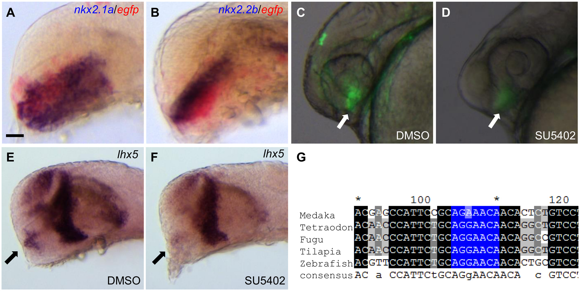
Fig. 3 CNS2 contains hypothalamic enhancer activity and responses to FGF signaling.
(A-B) Double in situ hybridization results indicate CNS2 contains hypothalamic enhancer activity. The hypothalamic marker nkx2.1a and nkx2.2b are stained in dark blue, reporter egfp stained in red. (C-D) SU5402 treatment severely reduces CNS2 activity. Vehicle DMSO treated embryos show restricted hypothalamic EGFP reporter expression (pointed by the arrow in C). Embryos treated with the FGF signaling inhibitor SU5402 during the segmentation stage (10-24hpf) show minimal EGFP signals in the hypothalamic region (arrow in D, n = 48/55). (E-F) SU5402 treatment down-regulates endogenous lhx5 expression in the hypothalamic region. Endogenous lhx5 shows robust expression in the hypothalamic region (pointed by the arrow in E). SU5402 treatment during the segmentation stage down-regulates lhx5 expression in the hypothalamic region (arrow in F, n = 25/28). (G) Multiple sequence alignments of the CNS2 region in the five teleost species. The identified FGF downstream factor Pea3 binding site is highlighted in blue. Lateral view of the forebrain regions of embryos at 24 hpf (A-F), anterior to the left. Scale bar: 40µm in A-B; 50µm in C-D.