
- Title
-
Vitamin K reduces hypermineralisation in zebrafish models of PXE and GACI
- Authors
- Mackay, E.W., Apschner, A., Schulte-Merker, S.
- Source
- Full text @ Development
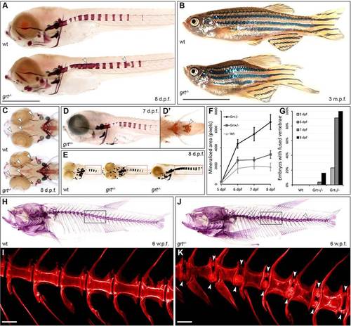
The gräte mutant phenotype is characterised by hypermineralisation in the skin and axial skeleton. (A) Alizarin Red staining of embryos at 8days post-fertilisation (dpf), demonstrating hypermineralisation along the vertebral column (arrowheads). Scale bar: 1mm. (B) Adult fish are viable, but feature a curved spine and reduced length. Scale bar: 1cm. (C) grt-/- embryos (ventral view) with enhanced mineralisation in craniofacial elements (arrowheads). (D) Skin mineralisation is infrequently seen in grt+/- and grt-/- embryos (D′, ventral view). (E) Representative images showing the area quantified by the mineralisation assay used in this and in subsequent figures. (F) Vertebral mineralisation in grt-/- embryos proceeds faster than in wild type, leading to (G) vertebral fusion from 6dpf onwards. (H,J) Alizarin Red staining at 6weeks post fertilisation (wpf) reveals a thickened, curved spine in grt-/- fish; confocal images (I,K) of the boxed regions reveal mineralised nodules on the margins of the intervertebral space (arrowheads). Scale bars: 0.1mm. |
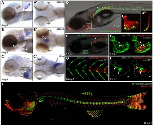
abcc6a is expressed at sites of mineralisation, but not the liver. (A) abcc6a transcripts are detected near the opercula (op) of 5dpf embryos. (B) abcc6b expression appears in the opercula (op), parasphenoid (ps), cleithrum (cl) and cartilage of the ear. Neither gene is detected in the liver, in contrast to (C) fetuin-A (ahsg). A,B,C, lateral views; A′,B′,C′, ventral views. (D) A transgenic reporter for abcc6a in a 7dpf embryo stained with Alizarin Red. GFP is seen in the notochord (arrowhead), operculum (D′) and cleithrum (D′′). (E,F) The abcc6a transgenic reporter in an embryo also expressing the osteoblast marker osterix:GFP, demonstrating abcc6a expression in some osteoblasts of the operculum. Expression can also be seen in the neural tube (arrowhead in E). (G) In juvenile (20dpf) vertebrae, abcc6a is expressed in the centra, whereas osx is expressed in the arches. (H) Some abcc6a+ osteoblasts also co-express the mature osteoblast marker osteocalcin:GFP. Dotted outline approximates the extent of the operculum at this stage. (I) In juvenile zebrafish, abcc6a is expressed in the intervertebral disc region, craniofacial bone elements and fins. Scale bars: 10µm in F,H. EXPRESSION / LABELING:
|
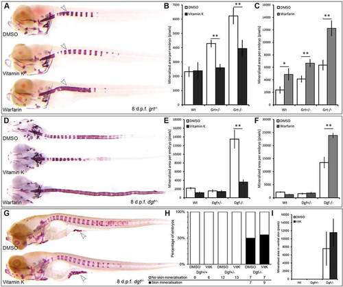
Vitamin K reduces hypermineralisation in gräte and dragonfish mutants. (A) Representative images of grt-/- embryos after treatment with vitamin K or warfarin from 4-8dpf, showing that vitamin K reduces hypermineralisation, whereas warfarin exacerbates it. (B,C) Quantification of Alizarin Red staining in A reveals significant rescue of the phenotype by vitamin K (n = 20 embryos per group). (D) Dgf-/- embryos treated with vitamin K or warfarin from 4-8dpf. (E,F) Quantification of Alizarin Red staining in D reveals a significant beneficial effect of vitamin K; results are similar to those seen in grt-/- embryos (n = 24 per group). (G) Alizarin Red staining of dgf-/- embryos exhibiting ectopic mineralisation in the ventral skin. Administration of vitamin K did not affect the incidence (H) or the extent (I) of this mineralisation. PHENOTYPE:
|
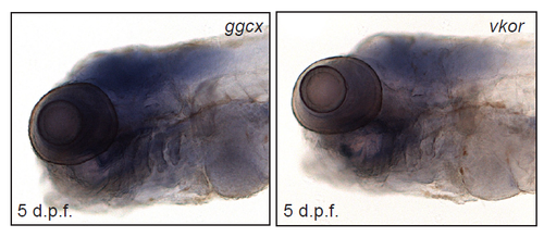
Whole-mount ISH for the vitamin K-metabolising enzymes, ggcx and vkor, indicate generalised expression in the head with no specific expression near the bone elements. |
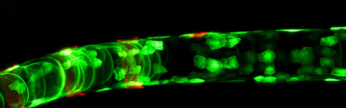
Expression of abcc6a in the notochord. GFP was observed in both vacuolated and notochord sheath cells; an example of the latter is indicated with an arrowhead. EXPRESSION / LABELING:
|