
Fig. 3
- ID
- ZDB-FIG-150422-42
- Publication
- Mackay et al., 2015 - Vitamin K reduces hypermineralisation in zebrafish models of PXE and GACI
- Other Figures
- All Figure Page
- Back to All Figure Page
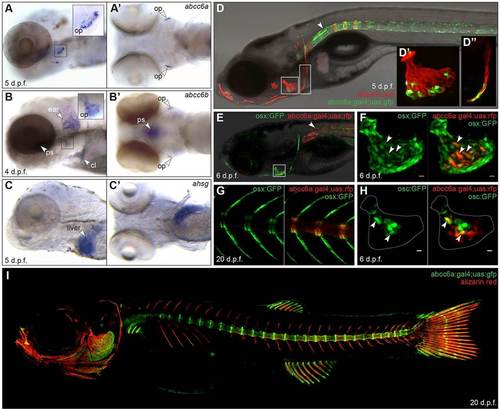
abcc6a is expressed at sites of mineralisation, but not the liver. (A) abcc6a transcripts are detected near the opercula (op) of 5dpf embryos. (B) abcc6b expression appears in the opercula (op), parasphenoid (ps), cleithrum (cl) and cartilage of the ear. Neither gene is detected in the liver, in contrast to (C) fetuin-A (ahsg). A,B,C, lateral views; A′,B′,C′, ventral views. (D) A transgenic reporter for abcc6a in a 7dpf embryo stained with Alizarin Red. GFP is seen in the notochord (arrowhead), operculum (D′) and cleithrum (D′′). (E,F) The abcc6a transgenic reporter in an embryo also expressing the osteoblast marker osterix:GFP, demonstrating abcc6a expression in some osteoblasts of the operculum. Expression can also be seen in the neural tube (arrowhead in E). (G) In juvenile (20dpf) vertebrae, abcc6a is expressed in the centra, whereas osx is expressed in the arches. (H) Some abcc6a+ osteoblasts also co-express the mature osteoblast marker osteocalcin:GFP. Dotted outline approximates the extent of the operculum at this stage. (I) In juvenile zebrafish, abcc6a is expressed in the intervertebral disc region, craniofacial bone elements and fins. Scale bars: 10µm in F,H. |
| Genes: | |
|---|---|
| Fish: | |
| Anatomical Terms: | |
| Stage Range: | Day 4 to Days 14-20 |