
- Title
-
Molecular dissection of Wnt3a-Frizzled8 interaction reveals essential and modulatory determinants of Wnt signaling activity
- Authors
- Kumar, S., Zigman, M., Patel, T.R., Trageser, B., Gross, J.C., Rahm, K., Boutros, M., Gradl, D., Steinbeisser, H., Holstein, T., Stetefeld, J., and Özbek, S.
- Source
- Full text @ BMC Biol.
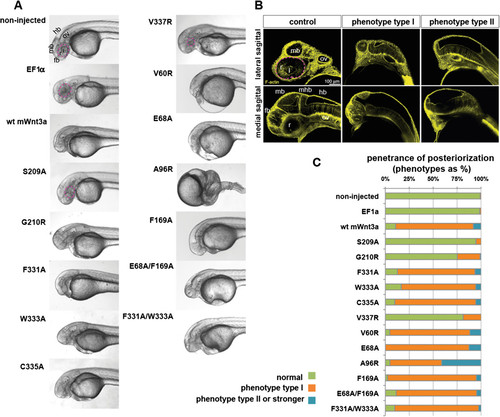
Expression of mouse Wnt3a mutants in zebrafish embryos. (A) Lateral view of zebrafish embryos, 1.5 days post fertilization (dpf), upon injection of capped mRNA encoding wild-type and mutant mouse Wnt3a capped mRNAs. Note lack of forebrain, eye field and midbrain structures in lateral view. (B) Posteriorizing effect of Wnt3a overexpression on zebrafish development as analyzed at 1.5 dpf. Pictures are single optical sections either of lateral (upper panel) or medial (lower panel) sagittal sections, with staining for F-actin marking cell outlines and thereby general nervous system morphology. The weaker phenotype with loss of retina and forebrain, though remnants of midbrain structures might be present, is termed phenotype type I. The stronger phenotype (lacking the forebrain, retina, lens and midbrain) is phenotype type II. (C) Penetrance of the dorsalized phenotype in zebrafish embryogenesis on wild-type and mutant mouse Wnt3a expression, quantified as a percentage. Type I (weaker) and type II (stronger) respond to the phenotypes shown in Figure 5B. Total number of embryos (n) from three independent experiments: non-injected: 403, EF1a: 76, wild-type mouse Wnt3a: 224, S209A: 113, G210R: 96, F331A: 79, W333A: 54, C335A: 91, V337R: 139, V60R: 86, E68A: 64, A96R: 124, F169A: 82, E68A/F169A: 88 and F331A/W333A: 82. In all panels, anterior is to the left and dorsal is up. Eyes are outlined with dashed pink lines. fb, forebrain; hb, hindbrain; l, lens; mb, midbrain; ov, otic vesicle; r, retina; WT, wild type; mhb, midbrain-hindbrain boundary. |

Influence of different V337 mutations on Wnt activity in vitro and in vivo. (A) Wnt reporter assay showing the influence of different amino acid side chains at position V337 on the signaling activity of Wnt3a. Position V337 is highly sensitive towards bulky side chains as illustrated in (B), which is an interaction model for the V337W mutant compared to the wild type. (C) Posteriorizing effect and (D) penetrance of the dorsalized phenotype in zebrafish embryogenesis upon expression of wild-type mouse Wnt3a and diverse V337 mutants. Experiments were carried out in triplicate. Error bars depict standard deviation. Statistical significance in relative luciferase activity levels compared to the wild-type Wnt3a levels in (A) as indicated: *P < 0.05, **P < 0.01, ***P < 0.001 and n.s. as not significant according to Student’s t-test. wt, wild type. |

Effect of ectopic mouse Wnt3a capped mRNA injections on zebrafish embryonic development. (A) Pictures of live embryos in lateral view. Anterior is to the left and posterior to the top. Note the effect of 3 pg mouse Wnt3a mRNA injection on loss of eye field and forebrain. Midbrain-hindbrain barrier (arrows) is still present. Upon injection of 33 pg mouse Wnt3a mRNA or more, the embryos are not able to gastrulate properly. (B) Quantification of the effect of full-length wild-type mouse Wnt3a mRNA on zebrafish embryonic development. Number of embryos analyzed for each condition: non-injected: n = 65, 3 pg: n = 81, 33 pg: n = 82, 100 pg: n = 93 and 150 pg: n = 74. (C) Levels of ectopic mouse Wnt3a protein expression in zebrafish embryos. The Western blot was probed with anti-His5 antibodies. It shows the total levels of His-tagged wild-type mouse Wnt3a and mutant proteins. The same membrane was probed with anti-alpha tubulin as a loading control. |