
Fig. 5
- ID
- ZDB-FIG-141007-87
- Publication
- Kumar et al., 2014 - Molecular dissection of Wnt3a-Frizzled8 interaction reveals essential and modulatory determinants of Wnt signaling activity
- Other Figures
- All Figure Page
- Back to All Figure Page
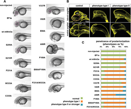
Expression of mouse Wnt3a mutants in zebrafish embryos. (A) Lateral view of zebrafish embryos, 1.5 days post fertilization (dpf), upon injection of capped mRNA encoding wild-type and mutant mouse Wnt3a capped mRNAs. Note lack of forebrain, eye field and midbrain structures in lateral view. (B) Posteriorizing effect of Wnt3a overexpression on zebrafish development as analyzed at 1.5 dpf. Pictures are single optical sections either of lateral (upper panel) or medial (lower panel) sagittal sections, with staining for F-actin marking cell outlines and thereby general nervous system morphology. The weaker phenotype with loss of retina and forebrain, though remnants of midbrain structures might be present, is termed phenotype type I. The stronger phenotype (lacking the forebrain, retina, lens and midbrain) is phenotype type II. (C) Penetrance of the dorsalized phenotype in zebrafish embryogenesis on wild-type and mutant mouse Wnt3a expression, quantified as a percentage. Type I (weaker) and type II (stronger) respond to the phenotypes shown in Figure 5B. Total number of embryos (n) from three independent experiments: non-injected: 403, EF1a: 76, wild-type mouse Wnt3a: 224, S209A: 113, G210R: 96, F331A: 79, W333A: 54, C335A: 91, V337R: 139, V60R: 86, E68A: 64, A96R: 124, F169A: 82, E68A/F169A: 88 and F331A/W333A: 82. In all panels, anterior is to the left and dorsal is up. Eyes are outlined with dashed pink lines. fb, forebrain; hb, hindbrain; l, lens; mb, midbrain; ov, otic vesicle; r, retina; WT, wild type; mhb, midbrain-hindbrain boundary. |