
- Title
-
Valproic Acid silencing of ascl1b/ascl1 results in the failure of serotonergic differentiation in a zebrafish model of Fetal Valproate Syndrome
- Authors
- Jacob, J., Ribes, V., Moore, S., Constable, S.C., Sasai, N., Gerety, S.S., Martin, D.J., Sergeant, C.P., Wilkinson, D.G., and Briscoe, J.
- Source
- Full text @ Dis. Model. Mech.
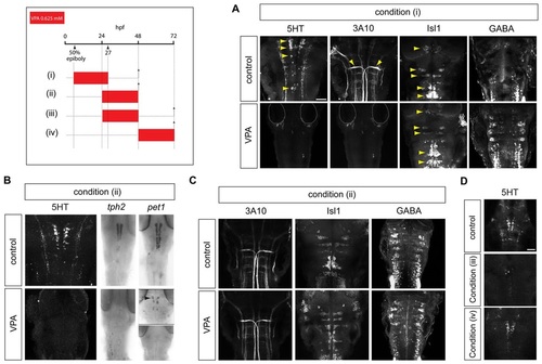
Effect of valproate exposure on the differentiation of serotonergic and other brainstem neuronal subtypes. The timing of drug treatment is indicated in this and all subsequent schematics by a coloured bar. Arrowheads mark the developmental stage of interest (hpf). Dashed lines with arrows indicate the developmental time point at which embryos were harvested. (A) Immunostaining for brainstem neuronal subtypes in 48 hpf zebrafish embryos exposed to VPA from 50% epiboly to 27 hpf [condition (i)] shows the failure of 5HT neuronal differentiation (n=22/22, P<0.0001), marked by 5HT (yellow arrowheads in control panel), and lack of Mauthner neurons (n=22/22), marked by the 3A10 anti-neurofilament monoclonal antibody (yellow arrowheads in control panel). Isl1+ motor neurons (yellow arrowheads in control and VPA panels) (n=10/10) and GABA-ergic neurons (n=10/10) persist. Scale bar: 50 µm. (B) Failure of serotonergic differentiation in embryos treated with VPA from 24-48 hpf [condition (ii)], as indicated by lack of 5HT (n=32/32, P<0.0001) and Tph2 expression. Pet1 (arrowhead) is severely reduced (n=3/15) or absent (n=12/15) in VPA-treated embryos. (C) Persistence of Mauthner, motor and GABA-ergic neurons in VPA treatment condition (ii). (D) At 72 hpf, embryos treated with VPA from 24-48 hpf [condition (iii)] show a limited recovery of 5HT expression (n=30/30). Treatment with VPA from 48-72 hpf [condition (iv)] does not affect serotonergic differentiation (n=30/30). hpf, hours post-fertilisation. Scale bar: 50 µm. EXPRESSION / LABELING:
PHENOTYPE:
|
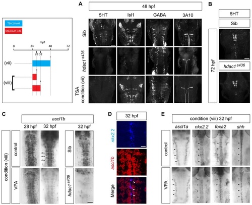
Failure of serotonergic differentiation in VPA-treated embryos is mimicked by blockade of Hdac1 activity and is associated with downregulation of ascl1b. (A) hdac1 mutation or pharmacological blockade mimics the effect of VPA on the differentiation of brainstem neuronal subtypes. At 48 hpf, hdac1s436 mutants lack 5HT neurons (n=15/15, P<0.0001) and show a severe depletion of Isl1+ motor neurons. Treatment with TSA from 24 to 48 hpf [condition (vii)] selectively affects 5HT expression (n=30/30, P<0.0001), but other neuronal subtypes are retained. Scale bar: 50 µm. (B) Partial recovery of 5HT neuronal differentiation in hdac1s436 mutants at 72 hpf (n=14/14). Scale bar: 50 µm. (C) Short duration (8 hour) exposure to VPA from 24 hpf [condition (vi)] leads to the downregulation of ascl1b expression (n=15/15), and similar loss of ascl1b expression is observed in hdac1s436 mutants (n=10/10). Rhombomeres (r) marked by arrowheads. Scale bar: 50 µm. (D) Double in situ hybridisation showing ascl1b expression in serotonergic progenitors, marked by expression of nkx2.2. Co-labelled cells are indicated by white arrowheads. Scale bar: 20 µm. (E) Short-duration VPA treatment [condition (viii)] does not affect the expression of the paralogous gene ascl1a, or other progenitor-expressed determinants of serotonergic fate, nkx2.2, foxa2 or shh (arrowheads). EXPRESSION / LABELING:
|
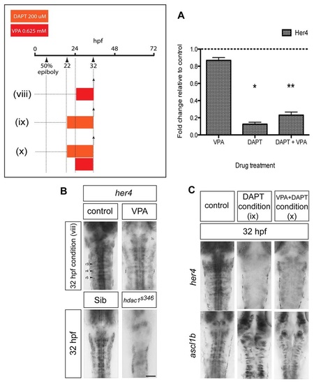
VPA treatment exposes cryptic transcriptional repression of ascl1b by basal levels of Notch signalling. (A) Quantitative reverse-transcriptase PCR showing that VPA mildly reduces her4 expression, whereas DAPT strongly reduces her4 expression to between 12% and 23% of control levels (*P=0.017, **P=0.001). (B) Short duration (8 hour) treatment with VPA [condition (viii)] does not upregulate her4 expression at 32 hpf (n=12/12). Downregulation of her4 expression in hdac1s436 mutant hindbrain at 32 hpf (n=5/5). Scale bar: 50 µm. (C) Concomitant blockade of Notch signalling using the γ-secretase inhibitor DAPT prevents the downregulation of ascl1b by VPA. Loss of her4 expression in DAPT-treated embryos [conditions (ix) and (x)] (upper panels) (n=35/35). Persistence of ascl1b expression in embryos treated with VPA and DAPT [condition (x)] (n=23/23). EXPRESSION / LABELING:
|
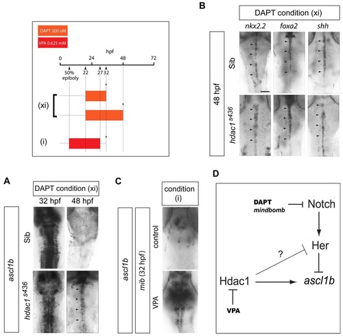
Ascl1b transcriptional regulation by opposing Hdac1 and Notch activities. (A) Blockade of Notch signalling by DAPT in hdac1s436 mutants [condition (xi)] results in the recovery of ascl1b expression at 32 hpf and 48 hpf (arrowheads) (n=15/15 for both developmental stages combined). Arrowheads indicate the expression domain of ascl1b. (B) DAPT treatment of hdac1s436 mutants [condition (xi)] does not affect the expression of nkx2.2 (n=7/7), foxa2 (n=8/8) and shh (n=6/6) (arrowheads) at 48 hpf. Arrowheads indicate the expression domain of the respective genes in the ventral midline of the embryo. Scale bar: 50 µm. (C) VPA treatment of embryos on the Notch signalling mutant mindbomb (mib) background [condition (i)] is associated with recovery of ascl1b expression (n=11/11). (D) Model of transcriptional regulation of ascl1b by Hdac1 and the Notch pathway. Solid arrows indicate direct positive regulation. Lines with an orthogonal bar represent inhibition. The inhibitory arrow with a question mark above it, from Hdac1 to Her, takes account of the possibility that another Her gene (or another transcription factor of unknown identity) that represses ascl1b is in turn repressed by Hdac1. |

Ascl1b is sufficient to rescue serotonergic differentiation in VPA-treated embryos. (A) Mis-expression of Myc-tagged Ascl1b in VPA-treated transgenic embryos rescues 5HT neuronal differentiation. A stable transgenic line expressing ERT2-GAL4 under the control of the ubiquitin (ubi) promoter was injected at the one-cell stage with plasmid DNA encoding Ascl1b-Myc under the control of UAS. Embryos were treated with VPA with or without 4-hydroxytamoxifen (4-OHT) [condition (xii)]. Myc immunostaining in shown in green. The upper and lower left panels show low-power views (scale bar: 500 µm) of Myc-immunostained zebrafish embryos treated with VPA in the absence (upper panel) or presence (lower panel) of 4-OHT. Addition of 4-OHT leads to widespread expression of Ascl1b-Myc (bottom left panel). Panels to the right show high-power views (scale bar: 50 µm) of Myc (green) and 5HT (red) immunostaining in the hindbrain of transgenic embryos. Embryos treated with VPA alone fail to express 5HT (n=51/51). Addition of 4-OHT and induction of Ascl1b-Myc expression rescues 5HT expression in 8/51 VPA-treated embryos (P=0.0058). Upper right and bottom right panels are high-power (scale bar: 20 µm) views of the boxed areas in the panels immediately to the left. The bottom, middle panel inset shows a higher-power view of the 5HT-expressing cells in the boxed area. The bottom right panel shows merged channels representing Ascl1b-Myc (green) and 5HT (red) immunostaining. (B) In hdac1s436 mutants treated with DAPT [condition (xi)], there is a rescue of 5HT neuronal differentiation at 48 hpf (n=22/22, P<0.0001). Scale bar: 50 µm. (C) On the mib mutant background there is a recovery of 5HT neurons at 48 hpf in VPA-treated embryos [condition (i)] (n=12/12, P=0.0002). Inset shows a high-power image of the boxed region. EXPRESSION / LABELING:
|