
- Title
-
Amyloid-beta and APP Deficiencies Cause Severe Cerebrovascular Defects: Important Work for an Old Villain
- Authors
- Luna, S., Cameron, D.J., and Ethell, D.W.
- Source
- Full text @ PLoS One
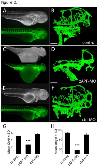
Cerebrovascular defects in APP-deficient zebrafish embryos. (A) Dark field (top) and fluorescence (bottom) images of a control transgenic embryo at 3 dpf shows vascular structures dues to EGFP expression in endothelial cells. (B) Confocal image (projected stack) of cerebrovascular structures in the head of the fish in A. (C) Dark field (top) and fluorescence (bottom) images of a zAPP-MO embryo at 3 dpf. (D) Confocal image (projected stack) of cerebrovascular structures in the head of the fish in C. (E) Dark field (top) and fluorescence (bottom) images of a ctrl-MO embryo at 3 dpf. (F) Confocal image (projected stack) of cerebrovascular structures in the head of the fish in E. (G) Graph showing the number of CtA branches in control (N = 30), zAPP-MO (N = 15), and ctrl-MO (N = 15) zebrafish at 3 dpf (***, P < 8.9e-16). (H) Mean CtA branch lengths in control (N = 28), zAPP-MO (N = 14), and ctrl-MO (N = 8) embryos at 3 dpf (***, P < 9.8e-23); scale bars = 100 μm. EXPRESSION / LABELING:
PHENOTYPE:
|
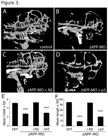
Aβ rescued vascular defects in APP-deficient (zAPP-MO) zebrafish embryos at 3 dpf. (A) Confocal image (projected stack) of a control zebrafish embryo at 3 dpf. (B) Comparable image of a zAPP-MO embryo at 3 dpf. (C) Cerebrovascular structures of an Aβ-treated zAPP-MO were similar to non-injected controls. (D) p3 treatment did not rescue vascular defects in zAPP-MO embryos. (E) Graph of CtA branch numbers in embryos in the control (N = 30), zAPP-MO (N = 15), Aβ-treated zAPP-MO (N = 15), and p3-treated zAPP-MO (N = 15) embryos at 3 dpf. Differences between control and zAPP-MO were significant (P < 8.9e-16), but there were no significant differences between Aβ-treated zAPP-MO and control or ctrl-MO. p3-treated zAPP-MO had significantly fewer branches than control embryos (P < 8.7e-14). (F) Graph of mean CtA branch lengths in control (N = 28), zAPP-MO (N = 14), Aβ-treated zAPP-MO (N = 10), and p3-treated zAPP-MO (N = 10) embryos at 3 dpf. Differences between control and zAPP-MO were significant (P < 9.8e-23), but there were no significant differences between Aβ-treated zAPP-MO and control or ctrl-MO. p3-treated zAPP-MO had significantly shorter vessel lengths than control embryos (P < 1.3e-15). EXPRESSION / LABELING:
PHENOTYPE:
|
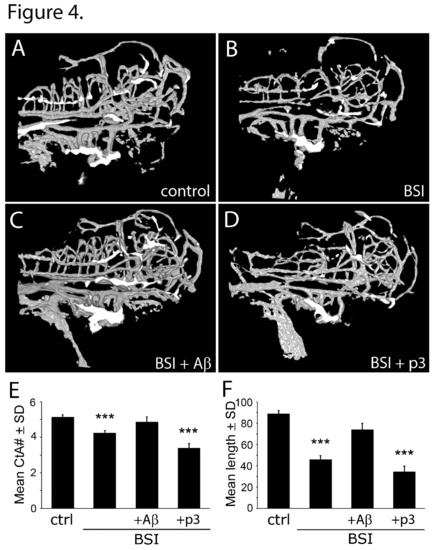
Aβ-deficiency induced by BSI-treatment caused vascular defects that were rescued by Aβ, but not p3.Vascular abnormalities in zAPP-MO could be discerned in embryos at 2 dpf. Fluorescence (top) and confocal (bottom) microscopy of 2 dpf embryos. (A) control uninjected embryo. (B) Embryo injected with scrambled sequence morpholino oligonucleotides (ctrl-MO). (C) Embryo injected with zAPP-targeting morpholino. Note CtA emerging from the lateral PHBC on the left side of A and B, but not C. EXPRESSION / LABELING:
|
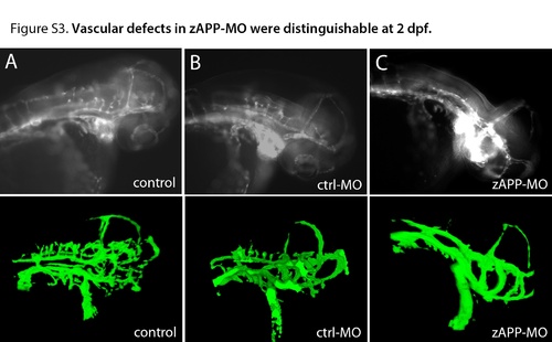
Vascular abnormalities in zAPP-MO could be discerned in embryos at 2 dpf. |