|
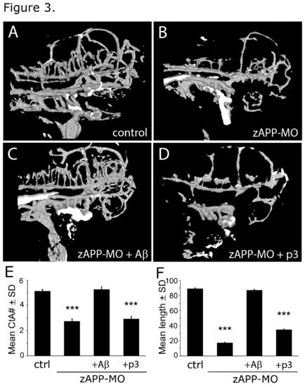
Aβ rescued vascular defects in APP-deficient (zAPP-MO) zebrafish embryos at 3 dpf. (A) Confocal image (projected stack) of a control zebrafish embryo at 3 dpf. (B) Comparable image of a zAPP-MO embryo at 3 dpf. (C) Cerebrovascular structures of an Aβ-treated zAPP-MO were similar to non-injected controls. (D) p3 treatment did not rescue vascular defects in zAPP-MO embryos. (E) Graph of CtA branch numbers in embryos in the control (N = 30), zAPP-MO (N = 15), Aβ-treated zAPP-MO (N = 15), and p3-treated zAPP-MO (N = 15) embryos at 3 dpf. Differences between control and zAPP-MO were significant (P < 8.9e-16), but there were no significant differences between Aβ-treated zAPP-MO and control or ctrl-MO. p3-treated zAPP-MO had significantly fewer branches than control embryos (P < 8.7e-14). (F) Graph of mean CtA branch lengths in control (N = 28), zAPP-MO (N = 14), Aβ-treated zAPP-MO (N = 10), and p3-treated zAPP-MO (N = 10) embryos at 3 dpf. Differences between control and zAPP-MO were significant (P < 9.8e-23), but there were no significant differences between Aβ-treated zAPP-MO and control or ctrl-MO. p3-treated zAPP-MO had significantly shorter vessel lengths than control embryos (P < 1.3e-15).
|