
IMAGE
Fig. S3
- ID
- ZDB-IMAGE-131231-13
- Publication
- Luna et al., 2013 - Amyloid-beta and APP Deficiencies Cause Severe Cerebrovascular Defects: Important Work for an Old Villain
- All Figures
- Figures for Luna et al., 2013
Image
|
Figure Caption
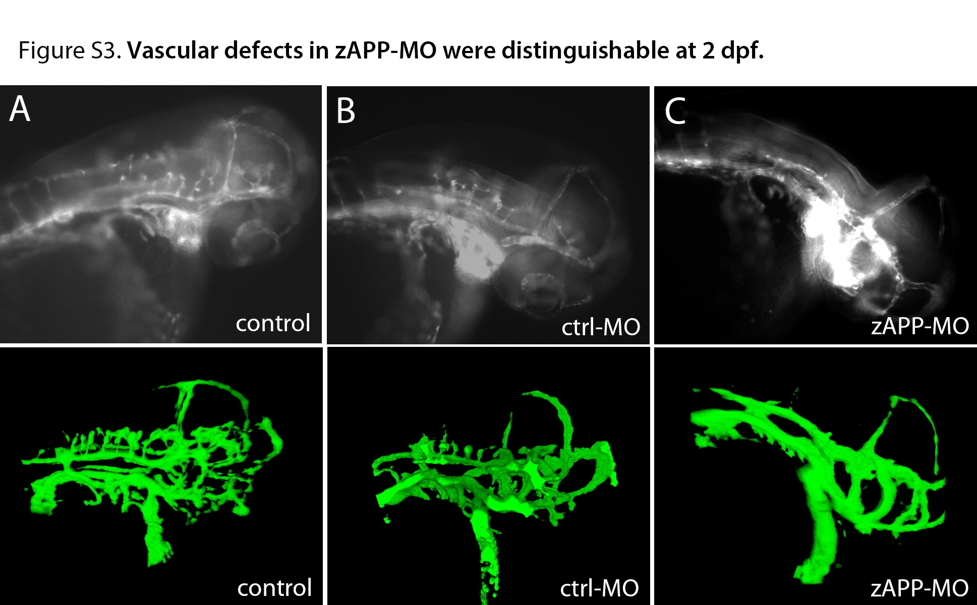
Fig. S3
Vascular abnormalities in zAPP-MO could be discerned in embryos at 2 dpf.
Fluorescence (top) and confocal (bottom) microscopy of 2 dpf embryos. (A) control uninjected embryo. (B) Embryo injected with scrambled sequence morpholino oligonucleotides (ctrl-MO). (C) Embryo injected with zAPP-targeting morpholino. Note CtA emerging from the lateral PHBC on the left side of A and B, but not C.
Acknowledgments
This image is the copyrighted work of the attributed author or publisher, and
ZFIN has permission only to display this image to its users.
Additional permissions should be obtained from the applicable author or publisher of the image.
Full text @ PLoS One