
- Title
-
Insulinoma-associated 1a (Insm1a) is required for photoreceptor differentiation in the zebrafish retina
- Authors
- Forbes-Osborne, M.A., Wilson, S.G., and Morris, A.C.
- Source
- Full text @ Dev. Biol.
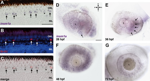
Insm1a is expressed in rod progenitor cells and in the developing retina. (A–C) Expression of insm1a in XOPS-mCFP retinas. (A) In situ hybridization with an antisense probe for insm1a was performed on retinal cryosections of XOPS-mCFP retinas after a 4 h exposure to BrdU. (B) Immunolabeling with anti-BrdU identified rod progenitor cells at the base of the ONL. (C) Overlay of in situ and immunolabeling demonstrates that many, but not all insm1a+ cells are also BrdU+. Arrows indicate insm1a+/BrdU+ cells; asterisks indicate insm1a+/BrdU cells. (D–G) Whole mount in situ hybridization of wild type embryos during development. (D) Insm1a expression was observed in the ventro-nasal patch between 24 and 28 hpf (arrow), (E) and insm1a expression expanded counterclockwise to the dorso-nasal retina at 36 hpf (arrows). (F) By 48 hpf, expression of insm1a had progressed to the dorso-temporal quadrant. (G) At 72 hpf, insm1a expression was only observed adjacent to the proliferative marginal zone at the retinal periphery. (A, scale bar=25 μm; D, scale bar=50 μm; D, dorsal; V, ventral; A, anterior; P, posterior; ONL, outer nuclear layer; INL, inner nuclear layer: L, lens; hpf, hours post fertilization.) |
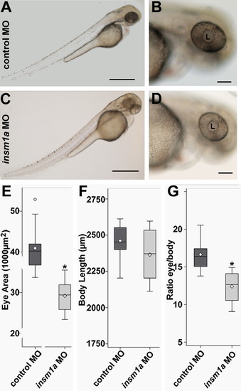
Insm1a-deficient embryos are microphthalmic. Compared to embryos injected with a control morpholino (A, B), insm1a morphants (C, D) displayed mild body torqueing and smaller eyes at 48 hpf. Whereas the eye area was significantly smaller in insm1a morphants (E), body lengths were not significantly different (F), and eye areas remained significantly smaller after controlling for body length (G). (A and C, scale bar=500 μm; B and D, scale bar=100 μm; E–G ⋄=mean; *p<0.0001, t-test; n>12; L, lens; hpf, hours post fertilization; MO, morpholino). PHENOTYPE:
|
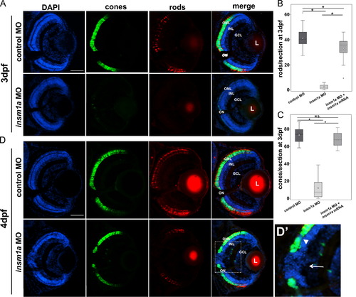
Photoreceptor cell differentiation is impaired in insm1a morphants. (A) At 3 dpf cone (green) and rod (red) photoreceptor cells were severely reduced in insm1a morphants. (B) The reduction in rods was significant and could be partially rescued by co-injection of in vitro transcribed insm1a mRNA with the insm1a morpholino (control: n=31 eyes from 22 embryos; insm1a MO: n=24 eyes from 17 embryos; insm1a MO+insm1a mRNA: n=18 eyes from 10 embryos). (C) The reduction in cones was significant and could be rescued by co-injection of in vitro transcribed insm1a mRNA with the insm1a morpholino (control: n=26 eyes from 17 embryos; insm1a MO: n=24 eyes from 17 embryos; insm1a MO+ insm1a mRNA n=18 eyes from 10 embryos). (D) At 4 dpf, the rod photoreceptors remain significantly reduced. However, cone photoreceptors (green) significantly recovered, but were less dense and contained visible gaps (dotted box). (D2) In regions lacking cones INL cells breached the OPL and extended into the ONL. Both small (arrowheads) and large (arrows) gaps were observed, accompanying the breached OPL. (B, D: ⋄=mean, *p<0.0001; scale bars=50 μm; ONL, outer nuclear layer; INL, inner nuclear layer; GCL, ganglion cell layer; L, lens; ON, optic nerve; dpf, days post fertilization; MO, morpholino). EXPRESSION / LABELING:
PHENOTYPE:
|
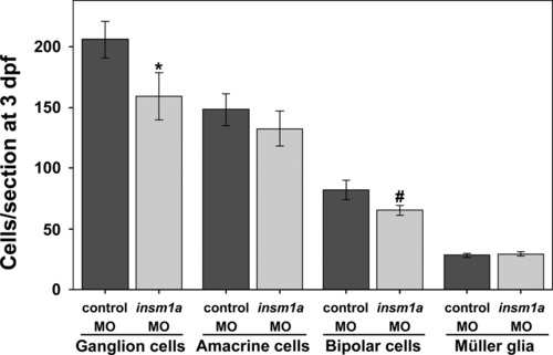
Knockdown of insm1a causes modest changes in cell number for some non-photoreceptor cell types. At 3 dpf the numbers of ganglion cells and bipolar cells were slightly reduced in insm1a morphant retinas, whereas the numbers of amacrine cells and Müller glia were not significantly affected. (*p<0.005; #p<0.003; mean±stdev for all; n=6 embryos each for ganglion and amacrine cells, 5 each for bipolar cells and 18 eyes from 10 embryos each for Müller glia). PHENOTYPE:
|
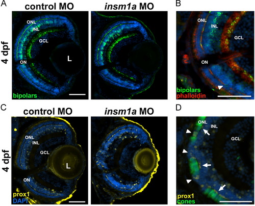
Bipolar and horizontal cell maturation is delayed in insm1a morphants. (A) At 4 dpf, bipolar cell morphology and labeling intensity in insm1a morphants appeared similar to controls. However, some breaches in the OPL were observed (B) and these regions (shown by an absence of phalloidin staining) occasionally contained bipolar cell bodies (white arrowhead). (C) At 4 dpf, Prox1+ cells were present in the INL adjacent to the OPL in insm1a morphant retinas; however, they were rounder and more widely spaced than in controls. (D) In regions of 4 dpf insm1a morphant retinas that contained cones, the adjacent Prox1+ horizontal cells displayed a more mature morphology (white arrows); however, in regions devoid of cones, the Prox1+ cells were absent or appeared less mature (white arrowheads). Prox1+ cells were also observed in the regions where cells from the INL had breached the ONL (white arrowheads). Scale bars=50 μm; ONL, outer nuclear layer; INL, inner nuclear layer; GCL, ganglion cell layer; IPL, inner plexiform layer; OPL, outer plexiform layer; L, lens; ON, optic nerve; dpf, days post fertilization; MO, morpholino. PHENOTYPE:
|
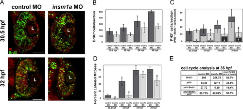
Cell cycle progression is delayed after insm1a knockdown. (A) At 30.5 hpf (0.5 hpi, top panels), BrdU+ cells (green) spanned the retina from basal to apical (dotted lines) in control retinas. However, in insm1a morphant retinas, the BrdU+ cells did not extend to the apical edge of the retina. At 32 hpf (2 hpi, bottom panels), fewer post-mitotic BrdU- cells were observed at the basal surface (dotted circles) in insm1a morphants compared with controls, indicating a delay in cell cycle exit in insm1a morphant retinas. (B) Quantitation of BrdU+ cells revealed a decrease in insm1a morphant retinas at all time points (*p<0.05). (C) The numbers of PH3+ cells were reduced at 2, 4 and 6 hpi in insm1a morphant retinas (*p<0.04). The number of cells double-positive for PH3 and BrdU was also reduced at all time points (#p<0.007). (D) The percent labeled mitoses (see Results for explanation) was significantly reduced in insm1a morphants at 0.5, 2 and 6 hpi, indicating that insm1a morphants took longer to progress from S phase into late G2/M phase compared with controls. (E) Summary of cell count analysis at 36 hpf (6 hpi); cell counts for insm1a morphants are expressed as a percentage of controls in the last column (for both controls and morphants: n=6 at 30.5, 32, and 34 hpf and n=3 at 36 hpf; mean±st.dev). Scale bars=50 μm; L, lens; PI, propidium iodide; hpf, hours post fertilization; hpi, hours post BrdU injection; MO, morpholino. PHENOTYPE:
|
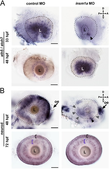
Insm1a acts upstream of pro-neural TFs ath5/atoh7 and neurod. (A) At 33 hpf ath5/atoh7 was expressed throughout the developing retina in controls; in contrast ath5/atoh7 expression was detectable only in the ventro-nasal patch of insm1a morphants (arrowhead). At 48 hpf, ath5/atoh7 expression in control retinas was restricted to the GCL; in insm1a morphant retinas, expression had expanded throughout the retina, in a pattern that closely resembled that of 33 hpf controls. (B) Neurod expression at 48 hpf was observed strongly in the developing photoreceptors and in the adjacent INL of control retinas. In insm1a morphants, however, neurod expression was mostly confined to the ventro-nasal retina (black arrowhead), with some expression throughout the central retina (gray arrowheads). In contrast, strong expression of neurod was observed in the olfactory epithelium (arrows) of insm1a morphants, which was absent in controls. By 72 hpf, neurod expression in the ONL (black bracket) and inner portion of the INL (blue bracket) was qualitatively similar in both controls and insm1a morphants. Scale bars=50 μm; D, dorsal; V, ventral; A, anterior; P, posterior; GCL, ganglion cell layer; oe, olfactory epithelium; ONL, outer nuclear layer INL, inner nuclear layer; hpf, hours post fertilization. EXPRESSION / LABELING:
|
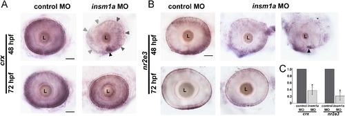
Expression of crx and nr2e3 is reduced at 48 hpf in insm1a morphants. (A) At 48 hpf, crx was expressed strongly in the developing ONL and weakly in some cells of the INL in control retinas. In insm1a morphant retinas crx was expressed in the ventro-nasal retina (black arrowhead), and expression decreased in a counterclockwise fashion across the retina (gray arrowheads). At 72 hpf, crx expression appeared similar between insm1a morphants and controls, and was observed primarily in the ONL. (B) At 48 hpf, nr2e3 was expressed strongly in nascent photoreceptor cells in the ONL, and in scattered cells in the distal half of INL in control retinas. Insm1a morphants displayed varying degrees of reduction in nr2e3 expression. In approximately half of the insm1a morphant retinas the nr2e3 expression pattern was only slightly less intense than controls (left panel), whereas the remaining half expressed nr2e3 only in the ventro-nasal retina (right, black arrowhead). At 72 hpf, nr2e3 expression had recovered greatly in insm1a morphants; however, nr2e3 staining did appear less dense and more discontinuous in insm1a morphants than in controls. (C) qPCR confirmed a 2.7-fold reduction in crx transcripts (*p<0.0128), and a 4.6-fold reduction in nr2e3 (*p<0.0084) at 48 hpf. Scale bar=50 μm; ONL, outer nuclear layer; INL, inner nuclear layer; L, lens; hpf, hours post fertilization; MO, morpholino. EXPRESSION / LABELING:
|

Notch-Delta and Her4 negatively regulate insm1a expression. (A, B) Pharmacologic inhibition of Notch signaling with DAPT resulted in an upregulation of insm1a expression in the developing eye and brain (B), compared to control (DMSO) treated embryos (A). (C) Co-transfection of HEK293 cells with a her4 cDNA expression vector repressed a luciferase reporter gene driven by either 300 bp (left) or 2.5 kb (right) of the insm1a promoter. D, dorsal; V, ventral; A, anterior; P, posterior; L, lens; hpf, hours post fertilization. EXPRESSION / LABELING:
|
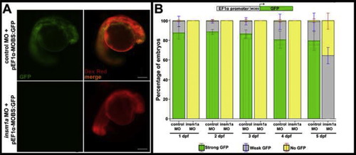
The insm1a MO is highly effective through 4 dpf. Embryos were co-injected with either standard control or insm1a MO and an eF1α-GFP plasmid containing the insm1a MO binding site (pEF1α-MOBS:GFP; n>40 embryos for each group). (A) At 1 dpf, control MO injected embryos showed robust, ubiquitous expression of GFP (top panels), whereas insm1a MO injected embryos showed no expression of GFP (bottom panels). Tetramethylrhodamine dextran (Dex Red) was used as an internal injection control. (B) Quantitation of the numbers of injected embryos showing GFP expression from 1 dpf through 5 dpf. GFP expression similar to that shown in (A) (top panel) was categorized as “strong GFP”. GFP expression that was limited to just a few cells in the embryo was categorized as “weak GFP” expression. “No GFP” required the complete absence of any identifiable GFP signal. From 1 dpf through 4 dpf, no insm1a morphants exhibited any GFP expression. In contrast, nearly all embryos co-injected with the standard control MO were GFP-positive, with over 90% strongly expressing GFP. By 5 dpf, nearly half of the insm1a morphants showed low levels of GFP expression, indicating a decline in the effectiveness of the insm1a MO. Scale bar=200 μm; MOBS, insm1a morpholino binding site; MO, morpholino; dpf, days post fertilization. |
Reprinted from Developmental Biology, 380(2), Forbes-Osborne, M.A., Wilson, S.G., and Morris, A.C., Insulinoma-associated 1a (Insm1a) is required for photoreceptor differentiation in the zebrafish retina, 157-171, Copyright (2013) with permission from Elsevier. Full text @ Dev. Biol.