
- Title
-
Epitope of titin a-band-specific monoclonal antibody Tit1 5 H1.1 is highly conserved in several Fn3 domains of the titin molecule. Centriole staining in human, mouse and zebrafish cells
- Authors
- Mikelsaar, A.V., Sünter, A., Mikelsaar, R., Toomik, P., Kõiveer, A., Mikelsaar, I., and Juronen, E.
- Source
- Full text @ Cell Div.
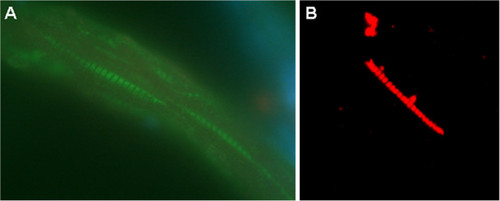
Immunohistochemical staining of skeletal muscle biopsy from the back side of zebrafish with MAb Tit1 5 H1.1. (A) Cryoslice of zebrafish skeletal muscle immunostained with MAb Tit1 5 H1.1, fixed with 4% PFA, specific staining (green) with Alexa 488, obj.100x. (B) Mechanically separated skeletal muscle fibre of zebrafish, fixed with 4% PFA, immunostained with MAb Tit1 5 H1.1, specific staining (red) with Alexa 594, obj.100x. Slices were embedded with Prolong Gold anti-fade reagent. EXPRESSION / LABELING:
|
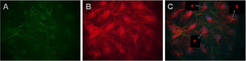
Immunofluorescence co-localization of MAb Tit1 5 H1.1 target antigen (titin) with F-actin in different cells of zebrafish primary culture of the testes. Cells were fixed on 3rd day of cultivation with 4% PFA, permeabilized, and double-labelled (C) with Mab Tit1 5 H1.1 for titin (red - Alexa 594) and with Phalloidin for F-actin (green – Alexa 488). Cell nuclei were stained blue with DAPI (obj. 100x). Arrows show centriole staining.In Photoshop image processing either red colour (A) or green colour (B) was removed and one can see independent staining both of F-actin (A) and titin (B). Note a very extensive fibrous centriole-orientated staining of titin. EXPRESSION / LABELING:
|
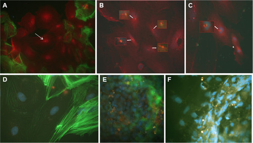
Co-location of MAb Tit1 5 H1.1 target antigen (titin) with F-actin and Î3-tubulin, negative staining with secondary antibodies,and the inhibition of specific staining of MAb Tit1 5 H1.1 with subpeptide N-AVNKYG-C in cells of zebrafish. (A) The cells of zebrafish testes were cultivated for 3 days, fixed, permeabilized, and double labeled with Alexa Fluor 488 conjugated to Phalloidin for F-actin (green) and with the MAb Tit1 5 H1.1 primary antibody for titin (specific staining with Alexa Fluor 594 conjugated to goat anti-mouse IgG secondary antibody (red). Arrow shows centriole staining. Note also a strong staining of nuclei and cytoplasm (obj. 40x). (B) The cells of zebrafish testes were double labeled with MAb Tit1 5 H1.1 for titin (red) and with rabbit anti-human Î3-tubulin for Î3-tubulin (green). Note the individual staining of the target antigens of both antibodies with centrioles/centrosomes. Arrows show centrosome/centriole staining.(C) Double labeling of mitotic cells of zebrafish with MAb Tit1 5 H1.1 for titin (red) and with rabbit anti-human Î3-tubulin for Î3-tubulin (green). The long arrow shows the co-location of MAb Tit1 5 H1.1 antigen (titin) and Î3-tubulin in the mitotic spindle with separated double-labeled centrioles (yellow stain), and the short arrow shows a late anaphase (only titin is labelled, red stain).(D) Negative staining of cells with secondary antibodies. (E,F) The cells were stained with MAb Tit1 5 H1.1 for titin but inhibited by the subpeptide N-AVNKYG-C. Note a full absence of staining with Alexa 594. Cell nuclei were stained blue with DAPI (obj. 100x). |