
Fig. 5
- ID
- ZDB-FIG-130228-33
- Publication
- Mikelsaar et al., 2012 - Epitope of titin a-band-specific monoclonal antibody Tit1 5 H1.1 is highly conserved in several Fn3 domains of the titin molecule. Centriole staining in human, mouse and zebrafish cells
- Other Figures
- All Figure Page
- Back to All Figure Page
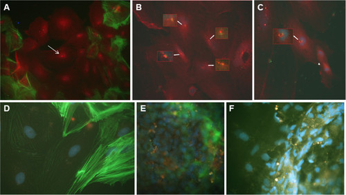
Co-location of MAb Tit1 5 H1.1 target antigen (titin) with F-actin and Î3-tubulin, negative staining with secondary antibodies,and the inhibition of specific staining of MAb Tit1 5 H1.1 with subpeptide N-AVNKYG-C in cells of zebrafish. (A) The cells of zebrafish testes were cultivated for 3 days, fixed, permeabilized, and double labeled with Alexa Fluor 488 conjugated to Phalloidin for F-actin (green) and with the MAb Tit1 5 H1.1 primary antibody for titin (specific staining with Alexa Fluor 594 conjugated to goat anti-mouse IgG secondary antibody (red). Arrow shows centriole staining. Note also a strong staining of nuclei and cytoplasm (obj. 40x). (B) The cells of zebrafish testes were double labeled with MAb Tit1 5 H1.1 for titin (red) and with rabbit anti-human Î3-tubulin for Î3-tubulin (green). Note the individual staining of the target antigens of both antibodies with centrioles/centrosomes. Arrows show centrosome/centriole staining.(C) Double labeling of mitotic cells of zebrafish with MAb Tit1 5 H1.1 for titin (red) and with rabbit anti-human Î3-tubulin for Î3-tubulin (green). The long arrow shows the co-location of MAb Tit1 5 H1.1 antigen (titin) and Î3-tubulin in the mitotic spindle with separated double-labeled centrioles (yellow stain), and the short arrow shows a late anaphase (only titin is labelled, red stain).(D) Negative staining of cells with secondary antibodies. (E,F) The cells were stained with MAb Tit1 5 H1.1 for titin but inhibited by the subpeptide N-AVNKYG-C. Note a full absence of staining with Alexa 594. Cell nuclei were stained blue with DAPI (obj. 100x). |